Division of Plastic and Reconstructive Surgery and Department of Surgery, Keck School of Medicine of USC, Los Angeles, California, United States of America.
Departments of Pathology and Dermatology, Keck School of Medicine of USC, Los Angeles, California, United States of America.
PLoS One. 2020 Jan 10;15(1):e0227599. doi: 10.1371/journal.pone.0227599. eCollection 2020.
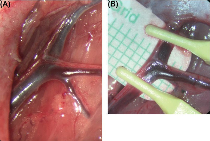
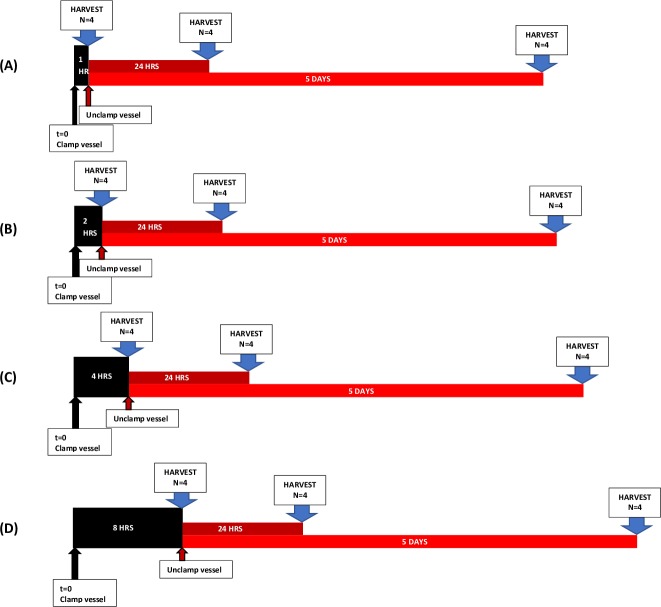
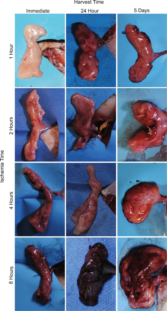
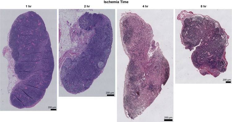
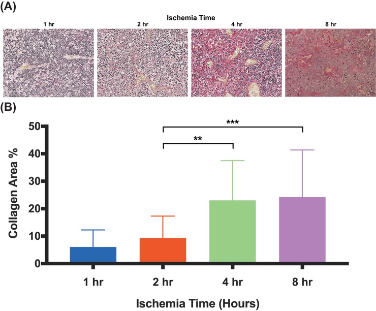
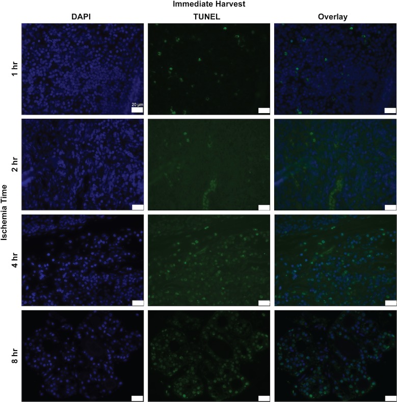
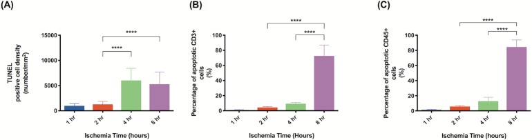
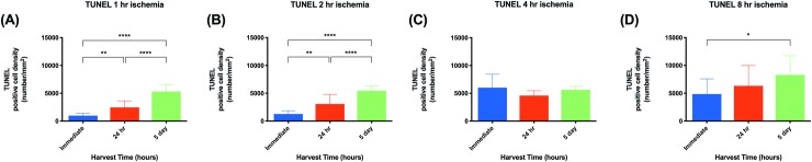
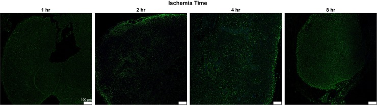
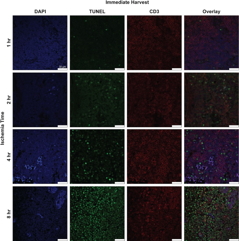
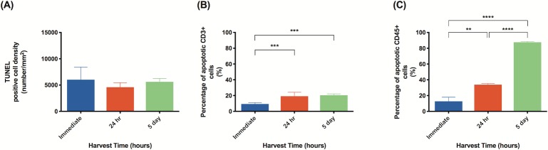
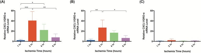
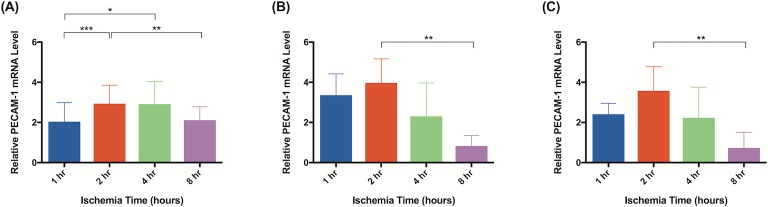
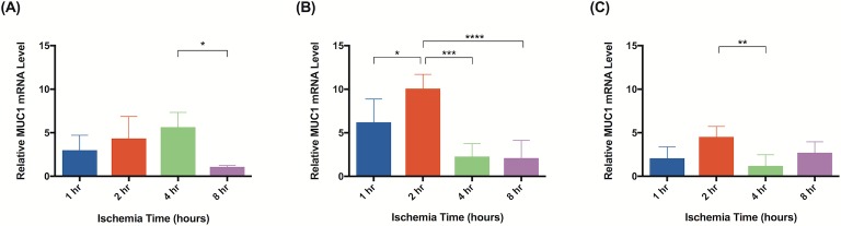
Vascularized lymph node transfer (VLNT) is a promising treatment modality for lymphedema; however, how lymphatic tissue responds to ischemia has not been well defined. This study investigates the cellular changes that occur in lymph nodes in response to ischemia and reperfusion. Lymph node containing superficial epigastric artery-based groin flaps were isolated in Prox-1 EGFP rats which permits real time identification of lymphatic tissue by green fluorescence during flap dissection. Flaps were subjected to ischemia for either 1, 2, 4, or 8 hours, by temporarily occluding the vascular pedicle. Flaps were harvested after 0 hours, 24 hours, or 5 days of reperfusion. Using EGFP signal guidance, lymph nodes were isolated from the flaps and tissue morphology, cell apoptosis, and inflammatory cytokines were quantified and analyzed via histology, immunostaining, and rtPCR. There was a significant increase in collagen deposition and tissue fibrosis in lymph nodes after 4 and 8 hours of ischemia compared to 1 and 2 hours, as assessed by picrosirius red staining. Cell apoptosis significantly increased after 4 hours of ischemia in all harvest times. In tissue subject to 4 hours of ischemia, longer reperfusion periods were associated with increased rates of CD3+ and CD45+ cell apoptosis. rtPCR analysis demonstrated significantly increased expression of CXCL1/GRO-α with 2 hours of ischemia and increased PECAM-1 and TNF-α expression with 1 hour of ischemia. Significant cell death and changes in tissue morphology do not occur until after 4 hours of ischemia; however, analysis of inflammatory biomarkers suggests that ischemia reperfusion injury can occur with as little as 2 hours of ischemia.
血管化淋巴结转移 (VLNT) 是治疗淋巴水肿的一种很有前途的治疗方法;然而,淋巴组织对缺血的反应尚未得到很好的定义。本研究调查了淋巴结在缺血再灌注过程中发生的细胞变化。在 Prox-1 EGFP 大鼠中分离含有腹壁浅动脉的腹股沟皮瓣的淋巴结,通过绿色荧光在皮瓣解剖过程中实时识别淋巴组织。通过暂时阻断血管蒂,将皮瓣缺血 1、2、4 或 8 小时。在再灌注 0 小时、24 小时或 5 天后收获皮瓣。使用 EGFP 信号引导,从皮瓣中分离出淋巴结,并通过组织形态学、细胞凋亡和炎症细胞因子的免疫染色和 rtPCR 定量和分析。与 1 小时和 2 小时相比,缺血 4 小时和 8 小时后,淋巴结中胶原沉积和组织纤维化显著增加,天狼星红染色评估。在所有收获时间中,缺血 4 小时后细胞凋亡明显增加。在组织缺血 4 小时的情况下,较长的再灌注时间与 CD3+和 CD45+细胞凋亡的增加率相关。rtPCR 分析表明,缺血 2 小时后 CXCL1/GRO-α 表达显著增加,缺血 1 小时后 PECAM-1 和 TNF-α 表达增加。直到缺血 4 小时后才会发生明显的细胞死亡和组织形态变化;然而,炎症生物标志物的分析表明,即使缺血 2 小时也可能发生缺血再灌注损伤。