• 文献检索
  • 文档翻译
  • 深度研究
  • 学术资讯
  • Suppr Zotero 插件Zotero 插件
  • 邀请有礼
  • 套餐&价格
  • 历史记录
应用&插件
Suppr Zotero 插件Zotero 插件浏览器插件Mac 客户端Windows 客户端微信小程序
定价
高级版会员购买积分包购买API积分包
服务
文献检索文档翻译深度研究API 文档MCP 服务
关于我们
关于 Suppr公司介绍联系我们用户协议隐私条款
关注我们

Suppr 超能文献

核心技术专利:CN118964589B侵权必究
粤ICP备2023148730 号-1Suppr @ 2026

文献检索

告别复杂PubMed语法,用中文像聊天一样搜索,搜遍4000万医学文献。AI智能推荐,让科研检索更轻松。

立即免费搜索

文件翻译

保留排版,准确专业,支持PDF/Word/PPT等文件格式,支持 12+语言互译。

免费翻译文档

深度研究

AI帮你快速写综述,25分钟生成高质量综述,智能提取关键信息,辅助科研写作。

立即免费体验

牙周炎患者上颌前牙牙槽骨的维度和形态:一项基于 CBCT 的回顾性分析。

The dimension and morphology of alveolar bone at maxillary anterior teeth in periodontitis: a retrospective analysis-using CBCT.

机构信息

Department of Periodontics, School of Stomatology, China Medical University, Shenyang, China.

出版信息

Int J Oral Sci. 2020 Jan 14;12(1):4. doi: 10.1038/s41368-019-0071-0.

DOI:10.1038/s41368-019-0071-0
PMID:31932579
原文链接:https://pmc.ncbi.nlm.nih.gov/articles/PMC6957679/
Abstract

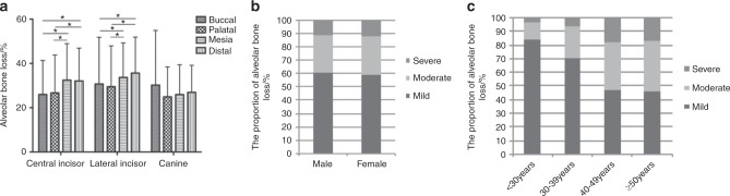
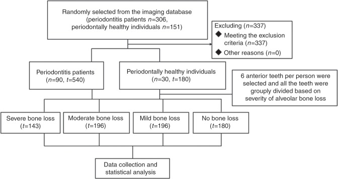
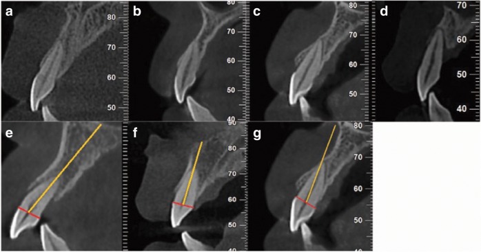
The morphology of the alveolar bone at the maxillary anterior teeth in periodontitis patients was evaluated by cone-beam computed tomography (CBCT) to investigate the distribution of alveolar defects and provide guidance for clinical practice. Ninety periodontitis patients and 30 periodontally healthy individuals were selected to determine the morphology of the alveolar bone at the maxillary anterior teeth according to the degree of bone loss, tooth type, sex and age. The differences in the dimensions between periodontitis patients and healthy individuals were compared, and the distribution of alveolar bone defects was analyzed. A classification system was established regarding the sagittal positions and angulations of the teeth. The buccal residual bone was thicker and the lingual bone was thinner in the periodontitis patients than in the periodontally healthy individuals, and there were differences between the different tooth types, sexes and age subgroups. The buccal undercut was close to the alveolar ridge, while fenestration was reduced and the apical bone height was higher in periodontitis patients than in periodontally healthy individuals. The apical bone height increased with the aggravation of bone loss and age. The proportions of different sagittal positions changed with the aggravation of bone loss. Moreover, the teeth moved more buccally regarding the positions of the maxillary anterior teeth. The morphology of the alveolar bone at the maxillary anterior teeth differed between periodontitis patients and healthy individuals, and the differences were related to the degree of bone loss, tooth type, sex and age.

摘要

通过锥形束 CT(CBCT)评估牙周炎患者上颌前牙牙槽骨的形态,以研究牙槽骨缺损的分布情况,为临床实践提供指导。选取 90 例牙周炎患者和 30 例牙周健康者,根据骨丧失程度、牙型、性别和年龄确定上颌前牙牙槽骨的形态。比较牙周炎患者与健康者之间各维度的差异,分析牙槽骨缺损的分布情况。建立了一种关于牙齿矢状位置和倾斜度的分类系统。与牙周健康者相比,牙周炎患者的颊侧剩余骨较厚,舌侧骨较薄,不同牙型、性别和年龄亚组之间存在差异。颊侧骨缺损接近牙槽嵴,而牙周炎患者的开窗减少,根尖骨高度增加。根尖骨高度随骨丧失和年龄的加重而增加。不同矢状位置的比例随骨丧失程度的加重而变化。此外,上颌前牙的位置移动更偏向颊侧。牙周炎患者和健康者上颌前牙牙槽骨形态不同,差异与骨丧失程度、牙型、性别和年龄有关。

https://cdn.ncbi.nlm.nih.gov/pmc/blobs/b2e5/6957679/ca805d8306d9/41368_2019_71_Fig5_HTML.jpg
https://cdn.ncbi.nlm.nih.gov/pmc/blobs/b2e5/6957679/4b93296081ba/41368_2019_71_Fig1_HTML.jpg
https://cdn.ncbi.nlm.nih.gov/pmc/blobs/b2e5/6957679/91f87c29fa85/41368_2019_71_Fig2_HTML.jpg
https://cdn.ncbi.nlm.nih.gov/pmc/blobs/b2e5/6957679/8b8d5649ad69/41368_2019_71_Fig3_HTML.jpg
https://cdn.ncbi.nlm.nih.gov/pmc/blobs/b2e5/6957679/61ee770645cd/41368_2019_71_Fig4_HTML.jpg
https://cdn.ncbi.nlm.nih.gov/pmc/blobs/b2e5/6957679/ca805d8306d9/41368_2019_71_Fig5_HTML.jpg

相似文献

1
The dimension and morphology of alveolar bone at maxillary anterior teeth in periodontitis: a retrospective analysis-using CBCT.牙周炎患者上颌前牙牙槽骨的维度和形态:一项基于 CBCT 的回顾性分析。
Int J Oral Sci. 2020 Jan 14;12(1):4. doi: 10.1038/s41368-019-0071-0.
2
Evaluation of buccal alveolar bone dimension of maxillary anterior and premolar teeth: a cone beam computed tomography investigation.上颌前牙和前磨牙颊牙槽骨尺寸的评估:锥形束 CT 研究。
Int J Oral Maxillofac Implants. 2012 Nov-Dec;27(6):1514-9.
3
[Investigation of sagittal root position in relation to the anterior maxillary alveolar bone: a cone-beam CT study in 300 cases with normal occlusion].[矢状面牙根位置与上颌前牙区牙槽骨关系的研究:300例正常咬合患者的锥形束CT研究]
Zhonghua Kou Qiang Yi Xue Za Zhi. 2017 Oct 9;52(10):631-636. doi: 10.3760/cma.j.issn.1002-0098.2017.10.010.
4
Anterior maxilla alveolar ridge dimension and morphology measurement by cone beam computerized tomography (CBCT) for immediate implant treatment planning.采用锥形束计算机断层扫描(CBCT)测量上颌前牙区牙槽嵴尺寸及形态,用于即刻种植治疗计划制定。
BMC Oral Health. 2015 Jun 10;15:65. doi: 10.1186/s12903-015-0055-1.
5
Assessment of gingival biotype and facial hard/soft tissue dimensions in the maxillary anterior teeth region using cone beam computed tomography.使用锥形束计算机断层扫描评估上颌前牙区牙龈生物型及面部软硬组织尺寸。
Arch Oral Biol. 2017 Jul;79:1-6. doi: 10.1016/j.archoralbio.2017.02.021. Epub 2017 Mar 1.
6
Analysis of facial bone wall dimensions and sagittal root position in the maxillary esthetic zone: a retrospective study using cone beam computed tomography.上颌美学区面部骨壁尺寸及牙根矢状位置分析:一项使用锥形束计算机断层扫描的回顾性研究
Int J Oral Maxillofac Implants. 2014 Sep-Oct;29(5):1123-9. doi: 10.11607/jomi.3348.
7
Correlation between buccal and alveolar bone widths at the central incisors according to cone-beam-computed tomography.根据锥形束计算机断层扫描评估中切牙颊侧与牙槽骨宽度之间的相关性。
Niger J Clin Pract. 2019 Jan;22(1):79-84. doi: 10.4103/njcp.njcp_320_18.
8
[Classification of the anatomical structures of maxillary central incisor root by cone-beam CT].[锥形束CT对上颌中切牙根解剖结构的分类]
Zhonghua Kou Qiang Yi Xue Za Zhi. 2017 Nov 9;52(11):656-660. doi: 10.3760/cma.j.issn.1002-0098.2017.11.002.
9
Three-dimensional evaluation of root dimensions and alveolar ridge width of maxillary lateral incisors in patients with unilateral agenesis.单侧牙缺失患者上颌侧切牙根尺寸和牙槽嵴宽度的三维评估
Prog Orthod. 2016 Dec;17(1):30. doi: 10.1186/s40510-016-0144-y. Epub 2016 Oct 10.
10
Cone beam computed tomographic measurement of maxillary central incisors to determine prevalence of facial alveolar bone width ≥2 mm.上颌中切牙锥形束 CT 测量用于确定面牙槽骨宽度≥2mm 的患病率。
Clin Implant Dent Relat Res. 2012 Aug;14(4):595-602. doi: 10.1111/j.1708-8208.2010.00287.x. Epub 2010 May 11.

引用本文的文献

1
Evaluating Factors Influencing Periodontal Bone Loss Using Cone Beam Computed Tomography: A Retrospective Study.使用锥形束计算机断层扫描评估影响牙周骨丧失的因素:一项回顾性研究
Med Sci Monit. 2025 May 2;31:e947759. doi: 10.12659/MSM.947759.
2
Expert consensus on orthodontic treatment of patients with periodontal disease.牙周病患者正畸治疗的专家共识
Int J Oral Sci. 2025 Apr 3;17(1):27. doi: 10.1038/s41368-025-00356-w.
3
Validation of novel measurement protocols proposed for the standardized assessment of crestal bone levels: A cone-beam computed tomography study.

本文引用的文献

1
Classification of sagittal root position in relation to the anterior maxillary osseous housing for immediate implant placement: a cone beam computed tomography study.矢状位根骨位置与即刻种植上前牙槽骨关系的分类:锥形束 CT 研究。
Int J Oral Maxillofac Implants. 2011 Jul-Aug;26(4):873-6.
2
Alveolar bone dehiscences and fenestrations: an anatomical study and review.牙槽骨缺损和开窗:一项解剖学研究及综述。
Rom J Morphol Embryol. 2009;50(3):391-7.
用于牙槽嵴骨水平标准化评估的新型测量方案的验证:一项锥形束计算机断层扫描研究。
Imaging Sci Dent. 2024 Dec;54(4):354-361. doi: 10.5624/isd.20240073. Epub 2024 Aug 12.
4
Advantages of using touch-controlled, minimally invasive implantation technique on soft tissue in the aesthetic zone of maxillary anterior teeth.在上颌前牙美学区软组织使用触控式微创植入技术的优势。
Medicine (Baltimore). 2024 Dec 13;103(50):e40051. doi: 10.1097/MD.0000000000040051.
5
Gingival spatial analysis reveals geographic immunological variation in a microbiota-dependent and -independent manner.牙龈空间分析揭示了微生物群依赖性和非依赖性的地理免疫学差异。
NPJ Biofilms Microbiomes. 2024 Dec 3;10(1):142. doi: 10.1038/s41522-024-00625-2.
6
Oro-Dental Characteristics in Patients With Adult-Onset Hypophosphatasia Compared to a Healthy Control Group-A Case-Control Study.成人起病型低磷酸酯酶症患者与健康对照组的口腔牙齿特征——一项病例对照研究
J Oral Rehabil. 2025 Jan;52(1):64-74. doi: 10.1111/joor.13878. Epub 2024 Oct 11.
7
Effects of Occlusal Contact on Maxillary Alveolar Bone Morphology in Patients with and without Anterior Open Bite: A Cross-Sectional Study.有无前牙开颌患者的咬合接触对上颌牙槽骨形态的影响:一项横断面研究
J Clin Med. 2024 May 23;13(11):3061. doi: 10.3390/jcm13113061.
8
Clinical Efficacy of Extracellular Vesicle Therapy in Periodontitis: Reduced Inflammation and Enhanced Regeneration.细胞外囊泡疗法在牙周炎中的临床疗效:减轻炎症和增强再生。
Int J Mol Sci. 2024 May 25;25(11):5753. doi: 10.3390/ijms25115753.
9
Prevalence and Severity of Circumferential Alveolar Bone Loss Using CBCT Images: A Retrospective Study of 20,620 Surfaces of 5155 Teeth.使用锥束计算机断层扫描(CBCT)图像评估圆周牙槽骨吸收的患病率和严重程度:一项对5155颗牙齿的20620个牙面的回顾性研究
Diagnostics (Basel). 2024 Feb 27;14(5):507. doi: 10.3390/diagnostics14050507.
10
Assessment of Posterior Maxillary Alveolar Bone for Immediate Implant Placement: A Quantitative and Qualitative Analysis.用于即刻种植的上颌后牙区牙槽骨评估:定量与定性分析
Eur J Dent. 2024 Jul;18(3):877-882. doi: 10.1055/s-0043-1777049. Epub 2024 Feb 8.