• 文献检索
  • 文档翻译
  • 深度研究
  • 学术资讯
  • Suppr Zotero 插件Zotero 插件
  • 邀请有礼
  • 套餐&价格
  • 历史记录
应用&插件
Suppr Zotero 插件Zotero 插件浏览器插件Mac 客户端Windows 客户端微信小程序
定价
高级版会员购买积分包购买API积分包
服务
文献检索文档翻译深度研究API 文档MCP 服务
关于我们
关于 Suppr公司介绍联系我们用户协议隐私条款
关注我们

Suppr 超能文献

核心技术专利:CN118964589B侵权必究
粤ICP备2023148730 号-1Suppr @ 2026

文献检索

告别复杂PubMed语法,用中文像聊天一样搜索,搜遍4000万医学文献。AI智能推荐,让科研检索更轻松。

立即免费搜索

文件翻译

保留排版,准确专业,支持PDF/Word/PPT等文件格式,支持 12+语言互译。

免费翻译文档

深度研究

AI帮你快速写综述,25分钟生成高质量综述,智能提取关键信息,辅助科研写作。

立即免费体验

脂肪干细胞衍生的细胞外囊泡通过内化诱导许旺细胞增殖。

Adipose Stem Cell-Derived Extracellular Vesicles Induce Proliferation of Schwann Cells via Internalization.

机构信息

Research Laboratory of the Division of Plastic and Reconstructive Surgery, Department of Surgery, Medical University of Vienna, 1090 Vienna, Austria.

出版信息

Cells. 2020 Jan 9;9(1):163. doi: 10.3390/cells9010163.

DOI:10.3390/cells9010163
PMID:31936601
原文链接:https://pmc.ncbi.nlm.nih.gov/articles/PMC7016740/
Abstract

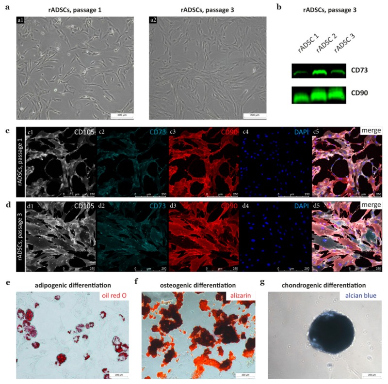
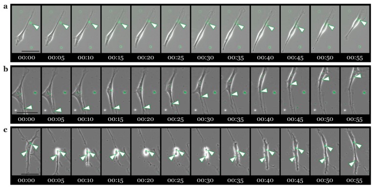
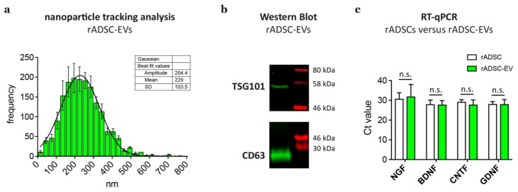
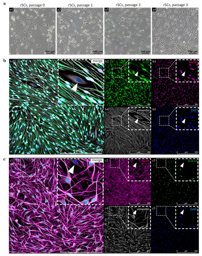
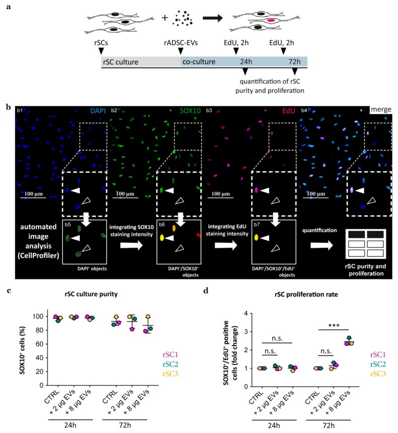
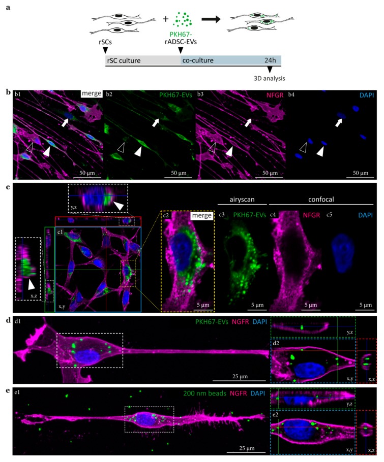
Recent studies showed a beneficial effect of adipose stem cell-derived extracellular vesicles (ADSC-EVs) on sciatic nerve repair, presumably through Schwann cell (SC) modulation. However, it has not yet been elucidated whether ADSC-EVs exert this supportive effect on SCs by extracellular receptor binding, fusion to the SC membrane, or endocytosis mediated internalization. ADSCs, ADSC-EVs, and SCs were isolated from rats and characterized according to associated marker expression and properties. The proliferation rate of SCs in response to ADSC-EVs was determined using a multicolor immunofluorescence staining panel followed by automated image analysis. SCs treated with ADSC-EVs and silica beads were further investigated by 3-D high resolution confocal microscopy and live cell imaging. Our findings demonstrated that ADSC-EVs significantly enhanced the proliferation of SCs in a time- and dose-dependent manner. 3-D image analysis revealed a perinuclear location of ADSC-EVs and their accumulation in vesicular-like structures within the SC cytoplasm. Upon comparing intracellular localization patterns of silica beads and ADSC-EVs in SCs, we found striking resemblance in size and distribution. Live cell imaging visualized that the uptake of ADSC-EVs preferentially took place at the SC processes from which the EVs were transported towards the nucleus. This study provided first evidence for an endocytosis mediated internalization of ADSC-EVs by SCs and underlines the therapeutic potential of ADSC-EVs in future approaches for nerve regeneration.

摘要

最近的研究表明脂肪干细胞衍生的细胞外囊泡 (ADSC-EVs) 对坐骨神经修复具有有益作用,可能通过施万细胞 (SC) 调节。然而,目前尚不清楚 ADSC-EVs 是否通过细胞外受体结合、融合到 SC 膜或内吞介导的内化来对 SC 发挥这种支持作用。从大鼠中分离 ADSC、ADSC-EVs 和 SC,并根据相关标志物表达和特性进行鉴定。通过多色免疫荧光染色面板和自动图像分析确定 SC 对 ADSC-EVs 的增殖率。用 ADSC-EVs 和硅胶珠处理 SC 后,通过 3-D 高分辨率共聚焦显微镜和活细胞成像进一步研究。我们的研究结果表明,ADSC-EVs 以时间和剂量依赖的方式显著增强了 SC 的增殖。3-D 图像分析显示 ADSC-EVs 位于核周位置,并在 SC 细胞质中的囊泡样结构中积累。在比较 SC 中硅胶珠和 ADSC-EVs 的细胞内定位模式时,我们发现它们在大小和分布上非常相似。活细胞成像可视化表明,ADSC-EVs 的摄取优先发生在 SC 过程中,EV 从这些过程中被运输到细胞核。这项研究首次提供了 ADSC-EVs 通过 SC 内吞摄取的证据,并强调了 ADSC-EVs 在未来神经再生方法中的治疗潜力。

https://cdn.ncbi.nlm.nih.gov/pmc/blobs/0e05/7016740/0d3cf5312d2f/cells-09-00163-g006.jpg
https://cdn.ncbi.nlm.nih.gov/pmc/blobs/0e05/7016740/5f85cd29588f/cells-09-00163-g001.jpg
https://cdn.ncbi.nlm.nih.gov/pmc/blobs/0e05/7016740/26d99aeb17ac/cells-09-00163-g002.jpg
https://cdn.ncbi.nlm.nih.gov/pmc/blobs/0e05/7016740/43be444ff9ed/cells-09-00163-g003.jpg
https://cdn.ncbi.nlm.nih.gov/pmc/blobs/0e05/7016740/2670bd324c96/cells-09-00163-g004.jpg
https://cdn.ncbi.nlm.nih.gov/pmc/blobs/0e05/7016740/e28b97156fb0/cells-09-00163-g005.jpg
https://cdn.ncbi.nlm.nih.gov/pmc/blobs/0e05/7016740/0d3cf5312d2f/cells-09-00163-g006.jpg

相似文献

1
Adipose Stem Cell-Derived Extracellular Vesicles Induce Proliferation of Schwann Cells via Internalization.脂肪干细胞衍生的细胞外囊泡通过内化诱导许旺细胞增殖。
Cells. 2020 Jan 9;9(1):163. doi: 10.3390/cells9010163.
2
Exosomes produced by adipose-derived stem cells inhibit schwann cells autophagy and promote the regeneration of the myelin sheath.脂肪来源干细胞分泌的外泌体抑制许旺细胞自噬,促进髓鞘再生。
Int J Biochem Cell Biol. 2021 Mar;132:105921. doi: 10.1016/j.biocel.2021.105921. Epub 2021 Jan 6.
3
Schwann cells secrete extracellular vesicles to promote and maintain the proliferation and multipotency of hDPCs.施万细胞分泌细胞外囊泡以促进和维持人牙髓细胞的增殖及多能性。
Cell Prolif. 2017 Aug;50(4). doi: 10.1111/cpr.12353.
4
Effect of exosomes from adipose-derived stem cells on the apoptosis of Schwann cells in peripheral nerve injury.脂肪来源干细胞外泌体对周围神经损伤雪旺细胞凋亡的影响。
CNS Neurosci Ther. 2020 Feb;26(2):189-196. doi: 10.1111/cns.13187. Epub 2019 Jul 6.
5
Delivery of miR-130a-3p Through Adipose-Derived Stem Cell-Secreted EVs Protects Against Diabetic Peripheral Neuropathy via DNMT1/NRF2/HIF1α/ACTA1 Axis.脂肪来源干细胞分泌的 EVs 通过 miR-130a-3p 传递来保护糖尿病周围神经病变,该途径涉及 DNMT1/NRF2/HIF1α/ACTA1 轴。
Mol Neurobiol. 2023 Jul;60(7):3678-3694. doi: 10.1007/s12035-023-03297-9. Epub 2023 Mar 18.
6
The Notch signalling pathway and miRNA regulation play important roles in the differentiation of Schwann cells from adipose-derived stem cells.Notch 信号通路和 miRNA 调控在脂肪来源干细胞向雪旺细胞分化中发挥重要作用。
Lab Invest. 2022 Mar;102(3):320-328. doi: 10.1038/s41374-021-00687-2. Epub 2021 Nov 18.
7
Schwann cell-like differentiated adipose stem cells promote neurite outgrowth via secreted exosomes and RNA transfer.许旺细胞样分化脂肪干细胞通过分泌的外泌体和 RNA 转移促进轴突生长。
Stem Cell Res Ther. 2018 Oct 11;9(1):266. doi: 10.1186/s13287-018-1017-8.
8
Schwann cell-derived EVs facilitate dental pulp regeneration through endogenous stem cell recruitment via SDF-1/CXCR4 axis.施万细胞衍生的 EV 通过 SDF-1/CXCR4 轴招募内源性干细胞促进牙髓再生。
Acta Biomater. 2022 Mar 1;140:610-624. doi: 10.1016/j.actbio.2021.11.039. Epub 2021 Nov 28.
9
Molecular and Cellular Mechanisms Involved in Adipose-derived stem cell and their extracellular vesicles in an Experimental Model of Cardio- renal Syndrome type 3: Histological and Biochemical Study.脂肪源性干细胞及其细胞外囊泡在 3 型心肾综合征实验模型中的分子和细胞机制:组织学和生化研究。
Tissue Cell. 2022 Aug;77:101842. doi: 10.1016/j.tice.2022.101842. Epub 2022 Jun 7.
10
Differential Therapeutic Effect of Extracellular Vesicles Derived by Bone Marrow and Adipose Mesenchymal Stem Cells on Wound Healing of Diabetic Ulcers and Correlation to Their Cargoes.骨髓间充质干细胞和脂肪间充质干细胞来源的细胞外囊泡对糖尿病溃疡伤口愈合的差异治疗作用及其与囊泡内容物的相关性
Int J Mol Sci. 2021 Apr 8;22(8):3851. doi: 10.3390/ijms22083851.

引用本文的文献

1
Exosome Therapy in Stress Urinary Incontinence: A Comprehensive Literature Review.应激性尿失禁的外泌体疗法:全面文献综述
Biomedicines. 2025 May 19;13(5):1229. doi: 10.3390/biomedicines13051229.
2
Indole-3-propionic acid promotes Schwann cell proliferation following peripheral nerve injury by activating the PI3K/AKT pathway.吲哚-3-丙酸通过激活PI3K/AKT信号通路促进周围神经损伤后雪旺细胞的增殖。
Neurotherapeutics. 2025 Mar 26:e00578. doi: 10.1016/j.neurot.2025.e00578.
3
Conditioning period impacts the morphology and proliferative effect of extracellular vesicles derived from rat adipose tissue derived stromal cell.

本文引用的文献

1
Exosomes from human adipose-derived stem cells promote sciatic nerve regeneration via optimizing Schwann cell function.人脂肪间充质干细胞来源的外泌体通过优化施万细胞功能促进坐骨神经再生。
J Cell Physiol. 2019 Dec;234(12):23097-23110. doi: 10.1002/jcp.28873. Epub 2019 May 23.
2
Intercellular Vesicular Transfer by Exosomes, Microparticles and Oncosomes - Implications for Cancer Biology and Treatments.外泌体、微颗粒和肿瘤小体介导的细胞间囊泡转运——对癌症生物学及治疗的影响
Front Oncol. 2019 Mar 6;9:125. doi: 10.3389/fonc.2019.00125. eCollection 2019.
3
Therapeutic Potential of Extracellular Vesicles for the Treatment of Nerve Disorders.
预处理期会影响源自大鼠脂肪组织间充质细胞的细胞外囊泡的形态和增殖效应。
J Nanobiotechnology. 2025 Mar 4;23(1):164. doi: 10.1186/s12951-025-03273-6.
4
Roles of extracellular vesicles from mesenchymal stem cells in regeneration.间充质干细胞来源的细胞外囊泡在再生中的作用。
Mol Cells. 2024 Dec;47(12):100151. doi: 10.1016/j.mocell.2024.100151. Epub 2024 Nov 13.
5
Activating PPARβ/δ-Mediated Fatty Acid β-Oxidation Mitigates Mitochondrial Dysfunction Co-induced by Environmentally Relevant Levels of Molybdenum and Cadmium in Duck Kidneys.激活过氧化物酶体增殖物激活受体β/δ介导的脂肪酸β氧化可减轻环境相关水平的钼和镉共同诱导的鸭肾线粒体功能障碍。
Biol Trace Elem Res. 2024 Nov 15. doi: 10.1007/s12011-024-04450-8.
6
Target specification and therapeutic potential of extracellular vesicles for regulating corneal angiogenesis, lymphangiogenesis, and nerve repair.用于调节角膜血管生成、淋巴管生成和神经修复的细胞外囊泡的靶点特性及治疗潜力
Ocul Surf. 2024 Oct;34:459-476. doi: 10.1016/j.jtos.2024.10.005. Epub 2024 Oct 18.
7
Nuclear Magnetic Resonance Treatment Induces ßNGF Release from Schwann Cells and Enhances the Neurite Growth of Dorsal Root Ganglion Neurons In Vitro.磁共振治疗诱导施万细胞释放 βNGF 并增强体外背根神经节神经元的轴突生长。
Cells. 2024 Sep 13;13(18):1544. doi: 10.3390/cells13181544.
8
Treating amyotrophic lateral sclerosis with allogeneic Schwann cell-derived exosomal vesicles: a case report.用异体雪旺细胞衍生的外泌体囊泡治疗肌萎缩侧索硬化症:一例报告。
Neural Regen Res. 2025 Apr 1;20(4):1207-1216. doi: 10.4103/NRR.NRR-D-23-01815. Epub 2024 Apr 3.
9
Recent advances in the use of extracellular vesicles from adipose-derived stem cells for regenerative medical therapeutics.脂肪来源干细胞细胞外囊泡在再生医学治疗中的应用的最新进展。
J Nanobiotechnology. 2024 Jun 6;22(1):316. doi: 10.1186/s12951-024-02603-4.
10
Mesenchymal stem cells' "garbage bags" at work: Treating radial nerve injury with mesenchymal stem cell-derived exosomes.间充质干细胞的“垃圾袋”在发挥作用:用间充质干细胞衍生的外泌体治疗桡神经损伤。
World J Stem Cells. 2024 May 26;16(5):467-478. doi: 10.4252/wjsc.v16.i5.467.
细胞外囊泡治疗神经疾病的潜力
Front Neurosci. 2019 Mar 5;13:163. doi: 10.3389/fnins.2019.00163. eCollection 2019.
4
The Success and Failure of the Schwann Cell Response to Nerve Injury.施万细胞对神经损伤反应的成功与失败
Front Cell Neurosci. 2019 Feb 11;13:33. doi: 10.3389/fncel.2019.00033. eCollection 2019.
5
Minimal information for studies of extracellular vesicles 2018 (MISEV2018): a position statement of the International Society for Extracellular Vesicles and update of the MISEV2014 guidelines.细胞外囊泡研究的最低限度信息2018(MISEV2018):国际细胞外囊泡协会的立场声明及MISEV2014指南的更新
J Extracell Vesicles. 2018 Nov 23;7(1):1535750. doi: 10.1080/20013078.2018.1535750. eCollection 2018.
6
Specificities of secretion and uptake of exosomes and other extracellular vesicles for cell-to-cell communication.细胞间通讯中外泌体和其他细胞外囊泡的分泌和摄取的特异性。
Nat Cell Biol. 2019 Jan;21(1):9-17. doi: 10.1038/s41556-018-0250-9. Epub 2019 Jan 2.
7
Imaging extracellular vesicles: current and emerging methods.细胞外囊泡的成像:当前和新兴的方法。
J Biomed Sci. 2018 Dec 24;25(1):91. doi: 10.1186/s12929-018-0494-5.
8
Bioactive Silk-Based Nerve Guidance Conduits for Augmenting Peripheral Nerve Repair.基于生物活性丝的神经引导导管在增强周围神经修复中的应用。
Adv Healthc Mater. 2018 Dec;7(23):e1800308. doi: 10.1002/adhm.201800308. Epub 2018 Sep 10.
9
Exosomes and Their MicroRNA Cargo: New Players in Peripheral Nerve Regeneration.外泌体及其 microRNA 货物:周围神经再生的新角色。
Neurorehabil Neural Repair. 2018 Sep;32(9):765-776. doi: 10.1177/1545968318798955.
10
CellProfiler 3.0: Next-generation image processing for biology.CellProfiler 3.0:生物学的下一代图像处理。
PLoS Biol. 2018 Jul 3;16(7):e2005970. doi: 10.1371/journal.pbio.2005970. eCollection 2018 Jul.