Beijing Tongren Eye Center, Beijing Tongren Hospital, Beijing Institute of Ophthalmology, Capital Medical University, Beijing, China.
Department of Ophthalmology, University of California, San Francisco School of Medicine, San Francisco, CA, United States of America.
PLoS One. 2020 Jan 28;15(1):e0227602. doi: 10.1371/journal.pone.0227602. eCollection 2020.
To provide in vivo measurements of anterior chamber angle structures and their relationship with age as evaluated by high-frequency ultrasound biomicroscopy (UBM) in patients with primary congenital glaucoma (PCG).
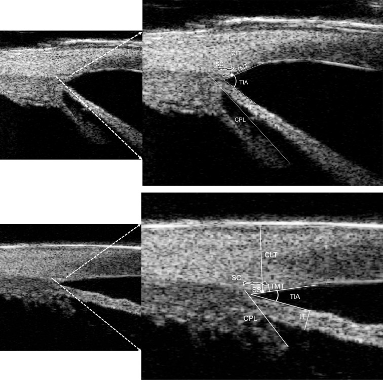
High-frequency UBM was done for 51 PCG eyes from 40 patients (aged from 3 to 96 months) and 11 unaffected contralateral eyes. Parameters, including the proportion of observable abnormal tissue membrane and Schlemm's canal, the largest cross-sectional area (CSA) of Schlemm's canal (SC), SC meridional diameter, trabecular-iris angle (TIA), trabecular meshwork (TM) thickness, iris thickness, ciliary process length, and corneal limbus thickness were compared between the two groups and their relationship with age was explored in PCG eyes.
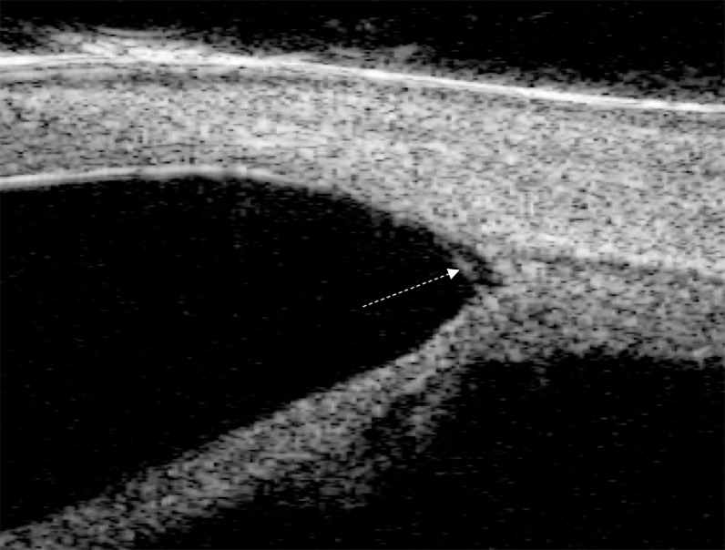
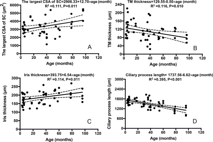
Abnormal tissue membrane was detected in 27.5% of PCG eyes and none in unaffected eyes. SC was observed in 73.1% of PGC eyes compared to 100% in unaffected eyes (P<0.001). The largest CSA of SC, SC meridional diameter, iris thickness, and corneal limbus thickness were all significantly smaller in PCG eyes compared to unaffected eyes (all P<0.05). TIA and ciliary process length in unaffected eyes were smaller than PCG eyes (both P<0.05). The largest CSA of SC, TM thickness, iris thickness, and ciliary process length were all significantly correlated to age in PCG eyes (P<0.05).
The anatomical information evaluated by high-frequency UBM may provide glaucoma specialists a useful tool to aid in understanding the dysgenesis and changes with age of anterior chamber angle in PCG.
通过高频超声生物显微镜(UBM)对原发性先天性青光眼(PCG)患者的前房角结构及其与年龄的关系进行活体测量。
对 40 例(年龄 3 至 96 个月)51 只 PCG 眼和 11 只对侧未受影响眼进行高频 UBM。比较两组患者的可观察到的异常组织膜和施累姆氏管的比例、施累姆氏管(SC)的最大横截面积(CSA)、SC 子午线直径、小梁虹膜角(TIA)、小梁网厚度、虹膜厚度、睫状体长度和角膜缘厚度等参数,并探讨 PCG 眼中这些参数与年龄的关系。
27.5%的 PCG 眼检测到异常组织膜,而未受影响眼则没有。73.1%的 PCG 眼观察到 SC,而未受影响眼为 100%(P<0.001)。与未受影响眼相比,PCG 眼的 SC 最大 CSA、SC 子午线直径、虹膜厚度和角膜缘厚度均显著较小(均 P<0.05)。未受影响眼的 TIA 和睫状体长度均小于 PCG 眼(均 P<0.05)。PCG 眼中的 SC 最大 CSA、TM 厚度、虹膜厚度和睫状体长度均与年龄显著相关(P<0.05)。
高频 UBM 评估的解剖学信息可为青光眼专家提供有用的工具,帮助了解 PCG 前房角的先天发育不良和随年龄变化。