• 文献检索
  • 文档翻译
  • 深度研究
  • 学术资讯
  • Suppr Zotero 插件Zotero 插件
  • 邀请有礼
  • 套餐&价格
  • 历史记录
应用&插件
Suppr Zotero 插件Zotero 插件浏览器插件Mac 客户端Windows 客户端微信小程序
定价
高级版会员购买积分包购买API积分包
服务
文献检索文档翻译深度研究API 文档MCP 服务
关于我们
关于 Suppr公司介绍联系我们用户协议隐私条款
关注我们

Suppr 超能文献

核心技术专利:CN118964589B侵权必究
粤ICP备2023148730 号-1Suppr @ 2026

文献检索

告别复杂PubMed语法,用中文像聊天一样搜索,搜遍4000万医学文献。AI智能推荐,让科研检索更轻松。

立即免费搜索

文件翻译

保留排版,准确专业,支持PDF/Word/PPT等文件格式,支持 12+语言互译。

免费翻译文档

深度研究

AI帮你快速写综述,25分钟生成高质量综述,智能提取关键信息,辅助科研写作。

立即免费体验

细胞激活过程中肝星状细胞外囊泡功能和蛋白质组组成的动态变化。

Dynamic Changes in Function and Proteomic Composition of Extracellular Vesicles from Hepatic Stellate Cells during Cellular Activation.

机构信息

Center for Clinical and Translational Research, The Abigail Wexner Research Institute, Nationwide Children's Hospital, Columbus, OH 43205, USA.

Department of Surgery, Wexner Medical Center, The Ohio State University, Columbus, OH 43210, USA.

出版信息

Cells. 2020 Jan 25;9(2):290. doi: 10.3390/cells9020290.

DOI:10.3390/cells9020290
PMID:31991791
原文链接:https://pmc.ncbi.nlm.nih.gov/articles/PMC7072607/
Abstract

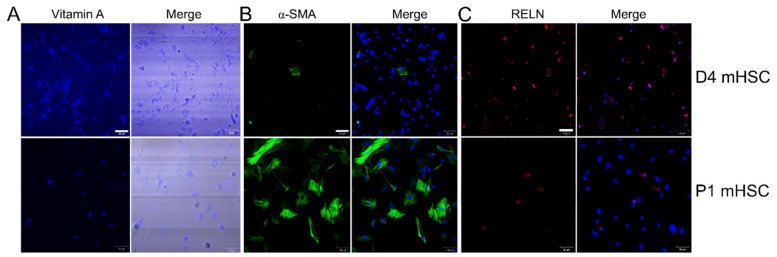
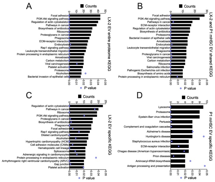
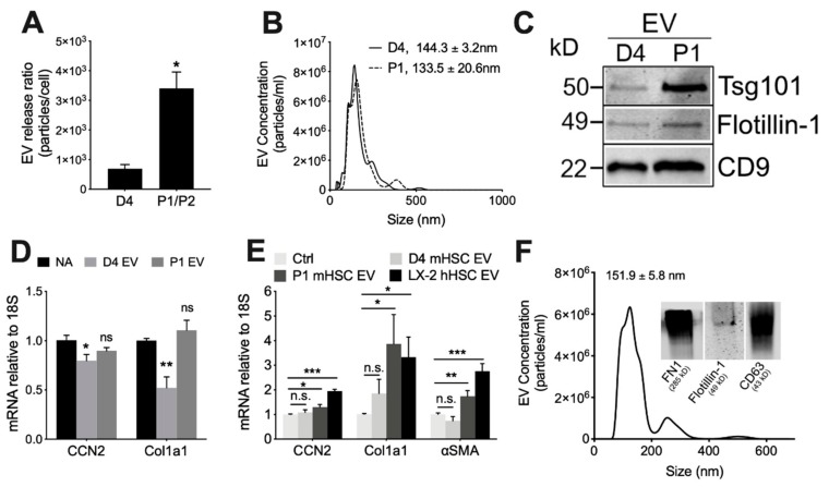
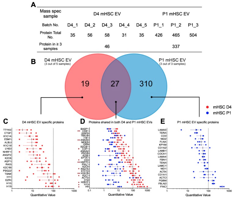
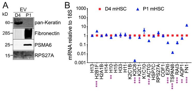
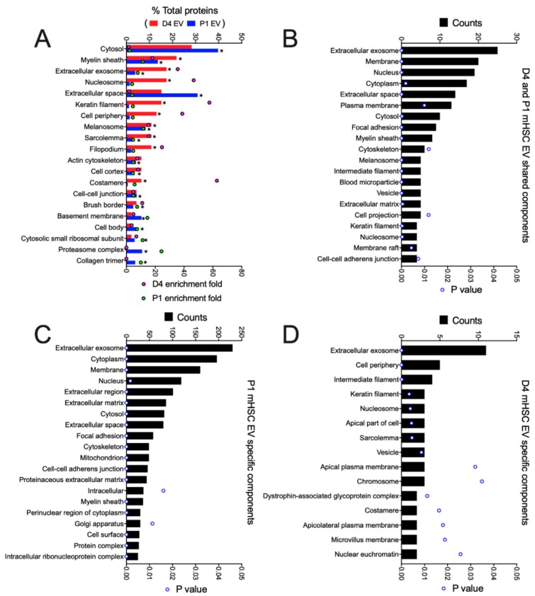
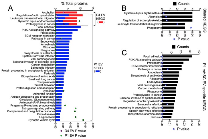
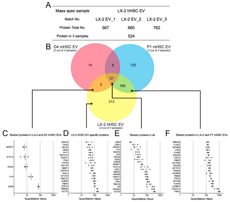
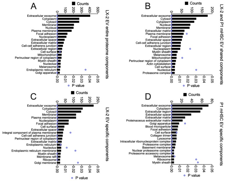
During chronic liver injury, hepatic stellate cells (HSC) undergo activation and are the principal cellular source of collagenous scar. In this study, we found that activation of mouse HSC (mHSC) was associated with a 4.5-fold increase in extracellular vesicle (EV) production and that fibrogenic gene expression (CCN2, Col1a1) was suppressed in Passage 1 (P1; activated) mHSC exposed to EVs from Day 4 (D4; relatively quiescent) mHSC but not to EVs from P1 mHSC. Conversely, gene expression (CCN2, Col1a1, αSMA) in D4 mHSC was stimulated by EVs from P1 mHSC but not by EVs from D4 mHSC. EVs from Day 4 mHSC contained only 46 proteins in which histones and keratins predominated, while EVs from P1 mHSC contained 337 proteins and these were principally associated with extracellular spaces or matrix, proteasome, collagens, vesicular transport, metabolic enzymes, ribosomes and chaperones. EVs from the activated LX-2 human HSC (hHSC) line also promoted fibrogenic gene expression in D4 mHSC in vitro and contained 524 proteins, many of which shared identity or had functional overlap with those in P1 mHSC EVs. The activation-associated changes in production, function and protein content of EVs from HSC likely contribute to the regulation of HSC function in vivo and to the fine-tuning of fibrogenic pathways in the liver.

摘要

在慢性肝损伤过程中,肝星状细胞(HSC)发生激活,是胶原性瘢痕的主要细胞来源。在这项研究中,我们发现小鼠 HSC(mHSC)的激活与细胞外囊泡(EV)产生增加 4.5 倍有关,并且纤维生成基因表达(CCN2、Col1a1)在第 1 代(激活)mHSC 暴露于第 4 天(相对静止)mHSC 的 EV 时受到抑制,但不被第 1 代 mHSC 的 EV 抑制。相反,第 4 天 mHSC 的基因表达(CCN2、Col1a1、αSMA)被第 1 代 mHSC 的 EV 刺激,但不受第 4 天 mHSC 的 EV 刺激。第 4 天 mHSC 的 EV 中仅包含 46 种蛋白质,其中组蛋白和角蛋白占主导地位,而第 1 代 mHSC 的 EV 中包含 337 种蛋白质,这些蛋白质主要与细胞外空间或基质、蛋白酶体、胶原蛋白、囊泡运输、代谢酶、核糖体和伴侣蛋白有关。来自激活的 LX-2 人 HSC(hHSC)系的 EV 也在体外促进第 4 天 mHSC 的纤维生成基因表达,并包含 524 种蛋白质,其中许多与第 1 代 mHSC EV 中的蛋白质具有相同的身份或功能重叠。HSC 来源的 EV 在产生、功能和蛋白质含量方面的激活相关变化可能有助于调节体内 HSC 功能,并精细调节肝脏中的纤维生成途径。

https://cdn.ncbi.nlm.nih.gov/pmc/blobs/4a1b/7072607/b2e41acb72c5/cells-09-00290-g009.jpg
https://cdn.ncbi.nlm.nih.gov/pmc/blobs/4a1b/7072607/20a078f32f45/cells-09-00290-g001.jpg
https://cdn.ncbi.nlm.nih.gov/pmc/blobs/4a1b/7072607/5b8c31cc5610/cells-09-00290-g002.jpg
https://cdn.ncbi.nlm.nih.gov/pmc/blobs/4a1b/7072607/b590429db2b8/cells-09-00290-g003.jpg
https://cdn.ncbi.nlm.nih.gov/pmc/blobs/4a1b/7072607/e55477e94754/cells-09-00290-g004.jpg
https://cdn.ncbi.nlm.nih.gov/pmc/blobs/4a1b/7072607/53cb9fb3f556/cells-09-00290-g005.jpg
https://cdn.ncbi.nlm.nih.gov/pmc/blobs/4a1b/7072607/4ffd91b97d37/cells-09-00290-g006.jpg
https://cdn.ncbi.nlm.nih.gov/pmc/blobs/4a1b/7072607/88dfcac990b1/cells-09-00290-g007.jpg
https://cdn.ncbi.nlm.nih.gov/pmc/blobs/4a1b/7072607/088fdcc9dd98/cells-09-00290-g008.jpg
https://cdn.ncbi.nlm.nih.gov/pmc/blobs/4a1b/7072607/b2e41acb72c5/cells-09-00290-g009.jpg

相似文献

1
Dynamic Changes in Function and Proteomic Composition of Extracellular Vesicles from Hepatic Stellate Cells during Cellular Activation.细胞激活过程中肝星状细胞外囊泡功能和蛋白质组组成的动态变化。
Cells. 2020 Jan 25;9(2):290. doi: 10.3390/cells9020290.
2
Hepatic stellate cell autophagy inhibits extracellular vesicle release to attenuate liver fibrosis.肝星状细胞自噬抑制细胞外囊泡释放以减轻肝纤维化。
J Hepatol. 2020 Nov;73(5):1144-1154. doi: 10.1016/j.jhep.2020.04.044. Epub 2020 May 8.
3
Human induced pluripotent stem cell-derived extracellular vesicles reduce hepatic stellate cell activation and liver fibrosis.人诱导多能干细胞衍生的细胞外囊泡可减少肝星状细胞的激活和肝纤维化。
JCI Insight. 2019 Jun 11;5(14):125652. doi: 10.1172/jci.insight.125652.
4
Suppression of fibrogenic signaling in hepatic stellate cells by Twist1-dependent microRNA-214 expression: Role of exosomes in horizontal transfer of Twist1.Twist1 依赖性微小RNA-214表达对肝星状细胞中纤维化信号的抑制作用:外泌体在Twist1水平转移中的作用
Am J Physiol Gastrointest Liver Physiol. 2015 Sep 15;309(6):G491-9. doi: 10.1152/ajpgi.00140.2015. Epub 2015 Jul 30.
5
Therapeutic effects of serum extracellular vesicles in liver fibrosis.血清细胞外囊泡在肝纤维化中的治疗作用。
J Extracell Vesicles. 2018 Apr 17;7(1):1461505. doi: 10.1080/20013078.2018.1461505. eCollection 2018.
6
Exosomes mediate intercellular transfer of pro-fibrogenic connective tissue growth factor (CCN2) between hepatic stellate cells, the principal fibrotic cells in the liver.外泌体介导肝星状细胞(肝脏主要的纤维化细胞)之间促纤维化的结缔组织生长因子(CCN2)的细胞间转移。
Surgery. 2014 Sep;156(3):548-55. doi: 10.1016/j.surg.2014.04.014. Epub 2014 Jun 2.
7
Epigenetic regulation of connective tissue growth factor by MicroRNA-214 delivery in exosomes from mouse or human hepatic stellate cells.外泌体来源的微小 RNA-214 介导的结缔组织生长因子的表观遗传调控作用:来自于鼠或人肝星状细胞。
Hepatology. 2014 Mar;59(3):1118-29. doi: 10.1002/hep.26768. Epub 2014 Jan 27.
8
Hepatic stellate cell-derived platelet-derived growth factor receptor-alpha-enriched extracellular vesicles promote liver fibrosis in mice through SHP2.肝星状细胞衍生的血小板衍生生长因子受体-α富集的细胞外囊泡通过 SHP2 促进小鼠肝纤维化。
Hepatology. 2018 Jul;68(1):333-348. doi: 10.1002/hep.29803. Epub 2018 May 10.
9
Extracellular Vesicles From Hepatocytes Are Therapeutic for Toxin-Mediated Fibrosis and Gene Expression in the Liver.肝细胞来源的细胞外囊泡对肝脏毒素介导的纤维化和基因表达具有治疗作用。
Front Cell Dev Biol. 2020 Jan 10;7:368. doi: 10.3389/fcell.2019.00368. eCollection 2019.
10
Fibrogenic Signaling Is Suppressed in Hepatic Stellate Cells through Targeting of Connective Tissue Growth Factor (CCN2) by Cellular or Exosomal MicroRNA-199a-5p.通过细胞或外泌体微小RNA-199a-5p靶向结缔组织生长因子(CCN2),肝星状细胞中的纤维化信号传导受到抑制。
Am J Pathol. 2016 Nov;186(11):2921-2933. doi: 10.1016/j.ajpath.2016.07.011. Epub 2016 Sep 21.

引用本文的文献

1
Therapeutic Actions of Hepatocyte Extracellular Vesicles in a Murine Model of Diet-Induced Steatohepatitis with Fibrosis.肝细胞外囊泡在饮食诱导的伴有纤维化的脂肪性肝炎小鼠模型中的治疗作用
Biomedicines. 2025 Jan 23;13(2):274. doi: 10.3390/biomedicines13020274.
2
Extracellular vesicle-mediated approaches for the diagnosis and therapy of MASLD: current advances and future prospective.细胞外囊泡介导的非酒精性脂肪性肝炎诊断与治疗方法:当前进展与未来展望
Lipids Health Dis. 2025 Jan 7;24(1):5. doi: 10.1186/s12944-024-02396-3.
3
Extracellular Vesicles, Circadian Rhythms, and Cancer: A Comprehensive Review with Emphasis on Hepatocellular Carcinoma.

本文引用的文献

1
Extracellular Vesicles From Hepatocytes Are Therapeutic for Toxin-Mediated Fibrosis and Gene Expression in the Liver.肝细胞来源的细胞外囊泡对肝脏毒素介导的纤维化和基因表达具有治疗作用。
Front Cell Dev Biol. 2020 Jan 10;7:368. doi: 10.3389/fcell.2019.00368. eCollection 2019.
2
HLSC-Derived Extracellular Vesicles Attenuate Liver Fibrosis and Inflammation in a Murine Model of Non-alcoholic Steatohepatitis.HLSC 衍生的细胞外囊泡可减轻非酒精性脂肪性肝炎小鼠模型的肝纤维化和炎症。
Mol Ther. 2020 Feb 5;28(2):479-489. doi: 10.1016/j.ymthe.2019.10.016. Epub 2019 Oct 28.
3
New and emerging anti-fibrotic therapeutics entering or already in clinical trials in chronic liver diseases.
细胞外囊泡、昼夜节律与癌症:一项重点关注肝细胞癌的综述
Cancers (Basel). 2024 Jul 16;16(14):2552. doi: 10.3390/cancers16142552.
4
Major vault protein is part of an extracellular cement material in the Atlantic salmon louse (Lepeophtheirus salmonis).主要穹窿蛋白是大西洋鲑虱(Lepeophtheirus salmonis)细胞外水泥物质的一部分。
Sci Rep. 2024 Jul 2;14(1):15240. doi: 10.1038/s41598-024-65683-0.
5
HepG2.2.15-derived exosomes facilitate the activation and fibrosis of hepatic stellate cells.HepG2.2.15 来源的外泌体促进肝星状细胞的激活和纤维化。
World J Gastroenterol. 2024 May 21;30(19):2553-2563. doi: 10.3748/wjg.v30.i19.2553.
6
Production, Exacerbating Effect, and EV-Mediated Transcription of Hepatic CCN2 in NASH: Implications for Diagnosis and Therapy of NASH Fibrosis.在 NASH 中 CCN2 的产生、加重作用和 EV 介导的转录:对 NASH 纤维化的诊断和治疗的影响。
Int J Mol Sci. 2023 Aug 15;24(16):12823. doi: 10.3390/ijms241612823.
7
Therapeutic role of FNDC5/irisin in attenuating liver fibrosis via inhibiting release of hepatic stellate cell-derived exosomes.FNDC5/鸢尾素通过抑制肝星状细胞衍生外泌体的释放减轻肝纤维化的治疗作用
Hepatol Int. 2023 Dec;17(6):1659-1671. doi: 10.1007/s12072-023-10523-y. Epub 2023 Apr 12.
8
Unraveling the Emerging Niche Role of Hepatic Stellate Cell-derived Exosomes in Liver Diseases.解析肝星状细胞衍生外泌体在肝脏疾病中新兴的特殊作用
J Clin Transl Hepatol. 2023 Apr 28;11(2):441-451. doi: 10.14218/JCTH.2022.00326. Epub 2022 Oct 18.
9
Pathophysiological mechanisms of hepatic stellate cells activation in liver fibrosis.肝纤维化中肝星状细胞激活的病理生理机制
World J Clin Cases. 2022 Apr 26;10(12):3662-3676. doi: 10.12998/wjcc.v10.i12.3662.
10
Autophagy and Exosomes: Cross-Regulated Pathways Playing Major Roles in Hepatic Stellate Cells Activation and Liver Fibrosis.自噬与外泌体:在肝星状细胞激活和肝纤维化中起主要作用的交叉调节途径
Front Physiol. 2022 Feb 3;12:801340. doi: 10.3389/fphys.2021.801340. eCollection 2021.
新型和新兴的抗纤维化治疗药物在慢性肝脏疾病中进入或已经进入临床试验。
Curr Opin Pharmacol. 2019 Dec;49:60-70. doi: 10.1016/j.coph.2019.09.006. Epub 2019 Oct 5.
4
Human induced pluripotent stem cell-derived extracellular vesicles reduce hepatic stellate cell activation and liver fibrosis.人诱导多能干细胞衍生的细胞外囊泡可减少肝星状细胞的激活和肝纤维化。
JCI Insight. 2019 Jun 11;5(14):125652. doi: 10.1172/jci.insight.125652.
5
Human bone marrow mesenchymal stem cells-derived exosomes alleviate liver fibrosis through the Wnt/β-catenin pathway.人骨髓间充质干细胞来源的外泌体通过 Wnt/β-catenin 通路缓解肝纤维化。
Stem Cell Res Ther. 2019 Mar 18;10(1):98. doi: 10.1186/s13287-019-1204-2.
6
Exosomal Transmission of MicroRNA from HCV Replicating Cells Stimulates Transdifferentiation in Hepatic Stellate Cells.来自丙型肝炎病毒复制细胞的微小RNA外泌体传递刺激肝星状细胞转分化。
Mol Ther Nucleic Acids. 2019 Mar 1;14:483-497. doi: 10.1016/j.omtn.2019.01.006. Epub 2019 Jan 18.
7
Extracellular Vesicles from Amnion-Derived Mesenchymal Stem Cells Ameliorate Hepatic Inflammation and Fibrosis in Rats.羊膜间充质干细胞来源的细胞外囊泡改善大鼠肝脏炎症和纤维化
Stem Cells Int. 2018 Dec 24;2018:3212643. doi: 10.1155/2018/3212643. eCollection 2018.
8
Exosomes Derived from Human Primed Mesenchymal Stem Cells Induce Mitosis and Potentiate Growth Factor Secretion.人诱导多能干细胞来源的外泌体诱导有丝分裂并增强生长因子分泌。
Stem Cells Dev. 2019 Mar 15;28(6):398-409. doi: 10.1089/scd.2018.0200. Epub 2019 Feb 26.
9
Minimal information for studies of extracellular vesicles 2018 (MISEV2018): a position statement of the International Society for Extracellular Vesicles and update of the MISEV2014 guidelines.细胞外囊泡研究的最低限度信息2018(MISEV2018):国际细胞外囊泡协会的立场声明及MISEV2014指南的更新
J Extracell Vesicles. 2018 Nov 23;7(1):1535750. doi: 10.1080/20013078.2018.1535750. eCollection 2018.
10
Hepatic fibrosis: It is time to go with hepatic stellate cell-specific therapeutic targets.肝纤维化:是时候针对肝星状细胞的特异性治疗靶点了。
Hepatobiliary Pancreat Dis Int. 2018 Jun;17(3):192-197. doi: 10.1016/j.hbpd.2018.04.003. Epub 2018 Apr 21.