• 文献检索
  • 文档翻译
  • 深度研究
  • 学术资讯
  • Suppr Zotero 插件Zotero 插件
  • 邀请有礼
  • 套餐&价格
  • 历史记录
应用&插件
Suppr Zotero 插件Zotero 插件浏览器插件Mac 客户端Windows 客户端微信小程序
定价
高级版会员购买积分包购买API积分包
服务
文献检索文档翻译深度研究API 文档MCP 服务
关于我们
关于 Suppr公司介绍联系我们用户协议隐私条款
关注我们

Suppr 超能文献

核心技术专利:CN118964589B侵权必究
粤ICP备2023148730 号-1Suppr @ 2026

文献检索

告别复杂PubMed语法,用中文像聊天一样搜索,搜遍4000万医学文献。AI智能推荐,让科研检索更轻松。

立即免费搜索

文件翻译

保留排版,准确专业,支持PDF/Word/PPT等文件格式,支持 12+语言互译。

免费翻译文档

深度研究

AI帮你快速写综述,25分钟生成高质量综述,智能提取关键信息,辅助科研写作。

立即免费体验

肝细胞来源的细胞外囊泡对肝脏毒素介导的纤维化和基因表达具有治疗作用。

Extracellular Vesicles From Hepatocytes Are Therapeutic for Toxin-Mediated Fibrosis and Gene Expression in the Liver.

作者信息

Li Xinlei, Chen Ruju, Kemper Sherri, Brigstock David R

机构信息

Center for Clinical and Translational Research, The Research Institute at Nationwide Children's Hospital, Columbus, OH, United States.

Department of Surgery, Wexner Medical Center, The Ohio State University, Columbus, OH, United States.

出版信息

Front Cell Dev Biol. 2020 Jan 10;7:368. doi: 10.3389/fcell.2019.00368. eCollection 2019.

DOI:10.3389/fcell.2019.00368
PMID:31998720
原文链接:https://pmc.ncbi.nlm.nih.gov/articles/PMC6966099/
Abstract

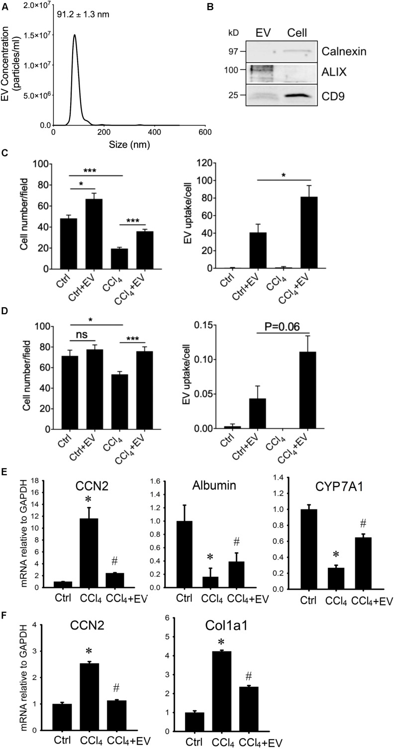
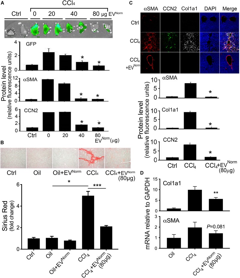
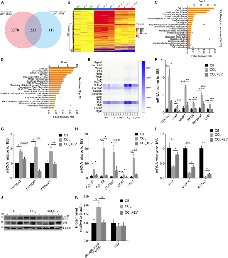
Extracellular vesicles (EVs) are nano-sized membrane-limited organelles that are liberated from their producer cells, traverse the intercellular space, and may interact with other cells resulting in the uptake of the EV molecular payload by the recipient cells which may become functionally reprogramed as a result. Previous studies showed that EVs purified from normal mouse AML12 hepatocytes ("EV") attenuate the pro-fibrogenic activities of activated hepatic stellate cells (HSCs), a principal fibrosis-producing cell type in the liver. In a 10-day CCl injury model, liver fibrogenesis, expression of hepatic cellular communication network factor 2 [CCN2, also known as connective tissue growth factor (CTGF)] or alpha smooth muscle actin (αSMA) was dose-dependently blocked during concurrent administration of EV. Hepatic inflammation and expression of inflammatory cytokines were also reduced by EV. In a 5-week CCl fibrosis model in mice, interstitial collagen deposition and mRNA and/or protein for collagen 1a1, αSMA or CCN2 were suppressed following administration of EV over the last 2 weeks. RNA sequencing (RNA-seq) revealed that EV therapy of mice receiving CCl for 5 weeks resulted in significant differences [false discovery rate (FDR) <0.05] in expression of 233 CCl-regulated hepatic genes and these were principally associated with fibrosis, cell cycle, cell division, signal transduction, extracellular matrix (ECM), heat shock, cytochromes, drug detoxification, adaptive immunity, and membrane trafficking. Selected gene candidates from these groups were verified by qRT-PCR as targets of EV in CCl-injured livers. Additionally, EV administration resulted in reduced activation of p53, a predicted upstream regulator of 40% of the genes for which expression was altered by EV following CCl liver injury. , EVs from human HepG2 hepatocytes suppressed fibrogenic gene expression in activated mouse HSC and reversed the reduced viability or proliferation of HepG2 cells or AML12 cells exposed to CCl. Similarly, EVs produced by primary human hepatocytes (PHH) protected PHH or human LX2 HSC from CCl-mediated changes in cell number or gene expression . These findings show that EVs from human or mouse hepatocytes regulate toxin-associated gene expression leading to therapeutic outcomes including suppression of fibrogenesis, hepatocyte damage, and/or inflammation.

摘要

细胞外囊泡(EVs)是纳米级的膜性细胞器,它们从产生细胞中释放出来,穿过细胞间空间,并可能与其他细胞相互作用,导致受体细胞摄取EV的分子载荷,受体细胞可能因此发生功能重编程。先前的研究表明,从正常小鼠AML12肝细胞中纯化的EVs(“EV”)可减弱活化的肝星状细胞(HSCs)的促纤维化活性,肝星状细胞是肝脏中主要的纤维化产生细胞类型。在为期10天的CCl损伤模型中,在同时给予EV期间,肝纤维化、肝细胞通讯网络因子2[CCN2,也称为结缔组织生长因子(CTGF)]或α平滑肌肌动蛋白(αSMA)的表达呈剂量依赖性被阻断。EV还可减轻肝脏炎症和炎性细胞因子的表达。在小鼠为期5周的CCl纤维化模型中,在最后2周给予EV后,间质胶原沉积以及胶原1a1、αSMA或CCN2的mRNA和/或蛋白表达均受到抑制。RNA测序(RNA-seq)显示,对接受CCl处理5周的小鼠进行EV治疗后,233个受CCl调节的肝脏基因的表达出现显著差异[错误发现率(FDR)<0.05],这些基因主要与纤维化、细胞周期、细胞分裂、信号转导、细胞外基质(ECM)、热休克、细胞色素、药物解毒、适应性免疫和膜运输有关。通过qRT-PCR验证了从这些组中选择的基因候选物是CCl损伤肝脏中EV的靶标。此外,给予EV导致p53的活化降低,p53是CCl肝损伤后其表达被EV改变的40%基因的预测上游调节因子。来自人HepG2肝细胞的EVs抑制活化的小鼠HSC中纤维化基因的表达,并逆转暴露于CCl的HepG2细胞或AML12细胞活力或增殖的降低。同样,原代人肝细胞(PHH)产生的EVs保护PHH或人LX2 HSC免受CCl介导的细胞数量或基因表达变化的影响。这些发现表明,来自人或小鼠肝细胞的EVs调节毒素相关基因的表达,从而产生包括抑制纤维化、肝细胞损伤和/或炎症在内的治疗效果。

https://cdn.ncbi.nlm.nih.gov/pmc/blobs/f6ad/6966099/4fa368dc3bbb/fcell-07-00368-g006.jpg
https://cdn.ncbi.nlm.nih.gov/pmc/blobs/f6ad/6966099/091b7b0b764a/fcell-07-00368-g001.jpg
https://cdn.ncbi.nlm.nih.gov/pmc/blobs/f6ad/6966099/5d851507125c/fcell-07-00368-g002.jpg
https://cdn.ncbi.nlm.nih.gov/pmc/blobs/f6ad/6966099/1bb09c82a771/fcell-07-00368-g003.jpg
https://cdn.ncbi.nlm.nih.gov/pmc/blobs/f6ad/6966099/bc49c86fa070/fcell-07-00368-g004.jpg
https://cdn.ncbi.nlm.nih.gov/pmc/blobs/f6ad/6966099/33ec1a8e0e32/fcell-07-00368-g005.jpg
https://cdn.ncbi.nlm.nih.gov/pmc/blobs/f6ad/6966099/4fa368dc3bbb/fcell-07-00368-g006.jpg

相似文献

1
Extracellular Vesicles From Hepatocytes Are Therapeutic for Toxin-Mediated Fibrosis and Gene Expression in the Liver.肝细胞来源的细胞外囊泡对肝脏毒素介导的纤维化和基因表达具有治疗作用。
Front Cell Dev Biol. 2020 Jan 10;7:368. doi: 10.3389/fcell.2019.00368. eCollection 2019.
2
Therapeutic effects of serum extracellular vesicles in liver fibrosis.血清细胞外囊泡在肝纤维化中的治疗作用。
J Extracell Vesicles. 2018 Apr 17;7(1):1461505. doi: 10.1080/20013078.2018.1461505. eCollection 2018.
3
Tetramethylpyrazine prevents liver fibrotic injury in mice by targeting hepatocyte-derived and mitochondrial DNA-enriched extracellular vesicles.川芎嗪通过靶向肝细胞来源的富含线粒体 DNA 的细胞外囊泡预防小鼠肝纤维化损伤。
Acta Pharmacol Sin. 2022 Aug;43(8):2026-2041. doi: 10.1038/s41401-021-00843-w. Epub 2022 Jan 13.
4
Hepatic stellate cell autophagy inhibits extracellular vesicle release to attenuate liver fibrosis.肝星状细胞自噬抑制细胞外囊泡释放以减轻肝纤维化。
J Hepatol. 2020 Nov;73(5):1144-1154. doi: 10.1016/j.jhep.2020.04.044. Epub 2020 May 8.
5
Structural and Functional Characterization of Fibronectin in Extracellular Vesicles From Hepatocytes.肝细胞外泌体中纤连蛋白的结构与功能表征
Front Cell Dev Biol. 2021 Mar 18;9:640667. doi: 10.3389/fcell.2021.640667. eCollection 2021.
6
Production, Exacerbating Effect, and EV-Mediated Transcription of Hepatic CCN2 in NASH: Implications for Diagnosis and Therapy of NASH Fibrosis.在 NASH 中 CCN2 的产生、加重作用和 EV 介导的转录:对 NASH 纤维化的诊断和治疗的影响。
Int J Mol Sci. 2023 Aug 15;24(16):12823. doi: 10.3390/ijms241612823.
7
Dynamic Changes in Function and Proteomic Composition of Extracellular Vesicles from Hepatic Stellate Cells during Cellular Activation.细胞激活过程中肝星状细胞外囊泡功能和蛋白质组组成的动态变化。
Cells. 2020 Jan 25;9(2):290. doi: 10.3390/cells9020290.
8
Extracellular Vesicular Transmission of miR-423-5p from HepG2 Cells Inhibits the Differentiation of Hepatic Stellate Cells.细胞外囊泡介导的 miR-423-5p 从 HepG2 细胞传递抑制肝星状细胞的分化。
Cells. 2022 May 23;11(10):1715. doi: 10.3390/cells11101715.
9
Aflatoxin B-exposed hepatocyte-derived extracellular vesicles: Initiating hepatic stellate cell-mediated liver fibrosis through a p53-Parkin-dependent mitophagy pathway.黄曲霉毒素 B 暴露的肝细胞衍生细胞外囊泡:通过 p53-Parkin 依赖的线粒体自噬途径引发肝星状细胞介导的肝纤维化。
Ecotoxicol Environ Saf. 2024 Jun 1;277:116363. doi: 10.1016/j.ecoenv.2024.116363. Epub 2024 Apr 25.
10
Human-Induced Hepatocytes-Derived Extracellular Vesicles Ameliorated Liver Fibrosis in Mice Via Suppression of TGF-β1/Smad Signaling and Activation of Nrf2/HO-1 Signaling.人诱导的肝细胞衍生细胞外囊泡通过抑制 TGF-β1/Smad 信号和激活 Nrf2/HO-1 信号改善小鼠肝纤维化。
Stem Cells Dev. 2023 Oct;32(19-20):638-651. doi: 10.1089/scd.2023.0110. Epub 2023 Jul 24.

引用本文的文献

1
Human Umbilical Cord Blood Plasma-Derived Exosomal miR-410-3p Alleviates Liver Injury by Regulating the Mitochondria-Mediated Antiapoptotic Signaling.人脐带血血浆来源的外泌体miR-410-3p通过调节线粒体介导的抗凋亡信号减轻肝损伤。
MedComm (2020). 2025 Aug 24;6(9):e70339. doi: 10.1002/mco2.70339. eCollection 2025 Sep.
2
Extracellular vesicles: emerging therapeutic agents for liver fibrosis.细胞外囊泡:肝脏纤维化的新兴治疗药物
Extracell Vesicles Circ Nucl Acids. 2025 May 7;6(2):216-244. doi: 10.20517/evcna.2025.08. eCollection 2025.
3
Therapeutic Actions of Hepatocyte Extracellular Vesicles in a Murine Model of Diet-Induced Steatohepatitis with Fibrosis.

本文引用的文献

1
HLSC-Derived Extracellular Vesicles Attenuate Liver Fibrosis and Inflammation in a Murine Model of Non-alcoholic Steatohepatitis.HLSC 衍生的细胞外囊泡可减轻非酒精性脂肪性肝炎小鼠模型的肝纤维化和炎症。
Mol Ther. 2020 Feb 5;28(2):479-489. doi: 10.1016/j.ymthe.2019.10.016. Epub 2019 Oct 28.
2
Human induced pluripotent stem cell-derived extracellular vesicles reduce hepatic stellate cell activation and liver fibrosis.人诱导多能干细胞衍生的细胞外囊泡可减少肝星状细胞的激活和肝纤维化。
JCI Insight. 2019 Jun 11;5(14):125652. doi: 10.1172/jci.insight.125652.
3
Changes of Circulating Extracellular Vesicles from the Liver after Roux-en-Y Bariatric Surgery.
肝细胞外囊泡在饮食诱导的伴有纤维化的脂肪性肝炎小鼠模型中的治疗作用
Biomedicines. 2025 Jan 23;13(2):274. doi: 10.3390/biomedicines13020274.
4
SHINE: SERS-based Hepatotoxicity detection using Inference from Nanoscale Extracellular vesicle content.SHINE:基于表面增强拉曼光谱的肝毒性检测,通过纳米级细胞外囊泡内容物推断得出
bioRxiv. 2025 Jan 30:2025.01.30.635446. doi: 10.1101/2025.01.30.635446.
5
Extracellular vesicles targeting non-parenchymal cells: the therapeutical effect on liver fibrosis.靶向非实质细胞的细胞外囊泡:对肝纤维化的治疗作用
eGastroenterology. 2024 Mar 19;2(1):e100040. doi: 10.1136/egastro-2023-100040. eCollection 2024 Jan.
6
Extracellular Vesicles in Viral Liver Diseases.细胞外囊泡在病毒性肝病中的作用。
Viruses. 2024 Nov 17;16(11):1785. doi: 10.3390/v16111785.
7
Extracellular Vesicles in Hepatobiliary Health and Disease.细胞外囊泡在肝胆健康与疾病中的作用
Compr Physiol. 2023 Jun 26;13(3):4631-4658. doi: 10.1002/cphy.c210046.
8
Liver Disease and Cell Therapy: Advances Made and Remaining Challenges.肝脏疾病与细胞治疗:进展与挑战
Stem Cells. 2023 Aug 16;41(8):739-761. doi: 10.1093/stmcls/sxad029.
9
Stem cell- derived extracellular vesicles as new tools in regenerative medicine - Immunomodulatory role and future perspectives.干细胞衍生的细胞外囊泡作为再生医学的新工具 - 免疫调节作用和未来展望。
Front Immunol. 2023 Jan 24;14:1120175. doi: 10.3389/fimmu.2023.1120175. eCollection 2023.
10
Native and engineered exosomes for inflammatory disease.用于炎症性疾病的天然和工程化外泌体。
Nano Res. 2023;16(5):6991-7006. doi: 10.1007/s12274-022-5275-5. Epub 2022 Dec 27.
Roux-en-Y 减肥手术后肝脏循环细胞外囊泡的变化。
Int J Mol Sci. 2019 Apr 30;20(9):2153. doi: 10.3390/ijms20092153.
4
Identification of key genes, pathways and potential therapeutic agents for liver fibrosis using an integrated bioinformatics analysis.使用综合生物信息学分析鉴定肝纤维化的关键基因、信号通路和潜在治疗药物。
PeerJ. 2019 Mar 22;7:e6645. doi: 10.7717/peerj.6645. eCollection 2019.
5
Human bone marrow mesenchymal stem cells-derived exosomes alleviate liver fibrosis through the Wnt/β-catenin pathway.人骨髓间充质干细胞来源的外泌体通过 Wnt/β-catenin 通路缓解肝纤维化。
Stem Cell Res Ther. 2019 Mar 18;10(1):98. doi: 10.1186/s13287-019-1204-2.
6
Extracellular Vesicles from Amnion-Derived Mesenchymal Stem Cells Ameliorate Hepatic Inflammation and Fibrosis in Rats.羊膜间充质干细胞来源的细胞外囊泡改善大鼠肝脏炎症和纤维化
Stem Cells Int. 2018 Dec 24;2018:3212643. doi: 10.1155/2018/3212643. eCollection 2018.
7
The official unified nomenclature adopted by the HGNC calls for the use of the acronyms, CCN1-6, and discontinuation in the use of CYR61, CTGF, NOV and WISP 1-3 respectively.HGNC采用的官方统一命名法要求使用首字母缩写CCN1 - 6,并分别停止使用CYR61、CTGF、NOV和WISP 1 - 3。
J Cell Commun Signal. 2018 Dec;12(4):625-629. doi: 10.1007/s12079-018-0491-1. Epub 2018 Nov 5.
8
The innate immune receptor TREM-1 promotes liver injury and fibrosis.天然免疫受体 TREM-1 促进肝损伤和纤维化。
J Clin Invest. 2018 Nov 1;128(11):4870-4883. doi: 10.1172/JCI98156. Epub 2018 Oct 2.
9
Therapeutic effects of serum extracellular vesicles in liver fibrosis.血清细胞外囊泡在肝纤维化中的治疗作用。
J Extracell Vesicles. 2018 Apr 17;7(1):1461505. doi: 10.1080/20013078.2018.1461505. eCollection 2018.
10
Human Umbilical Cord MSC-Derived Exosomes Suppress the Development of CCl-Induced Liver Injury through Antioxidant Effect.人脐带间充质干细胞来源的外泌体通过抗氧化作用抑制CCl诱导的肝损伤的发展。
Stem Cells Int. 2018 Mar 4;2018:6079642. doi: 10.1155/2018/6079642. eCollection 2018.