Department of Psychiatry, University of Cambridge, Cambridge CB2 0SZ, United Kingdom;
Department of Neuroimaging, Institute of Psychiatry, Psychology and Neurosciences, King's College London, London SE5 8AF, United Kingdom.
Proc Natl Acad Sci U S A. 2020 Feb 11;117(6):3248-3253. doi: 10.1073/pnas.1906144117. Epub 2020 Jan 28.
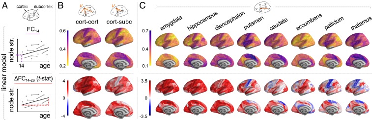
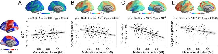
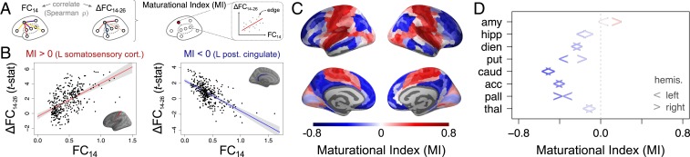
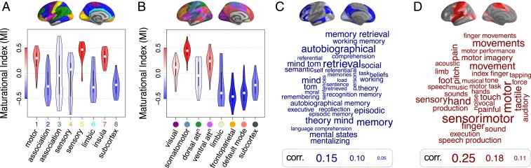
Adolescent changes in human brain function are not entirely understood. Here, we used multiecho functional MRI (fMRI) to measure developmental change in functional connectivity (FC) of resting-state oscillations between pairs of 330 cortical regions and 16 subcortical regions in 298 healthy adolescents scanned 520 times. Participants were aged 14 to 26 y and were scanned on 1 to 3 occasions at least 6 mo apart. We found 2 distinct modes of age-related change in FC: "conservative" and "disruptive." Conservative development was characteristic of primary cortex, which was strongly connected at 14 y and became even more connected in the period from 14 to 26 y. Disruptive development was characteristic of association cortex and subcortical regions, where connectivity was remodeled: connections that were weak at 14 y became stronger during adolescence, and connections that were strong at 14 y became weaker. These modes of development were quantified using the maturational index (MI), estimated as Spearman's correlation between edgewise baseline FC (at 14 y, [Formula: see text]) and adolescent change in FC ([Formula: see text]), at each region. Disruptive systems (with negative MI) were activated by social cognition and autobiographical memory tasks in prior fMRI data and significantly colocated with prior maps of aerobic glycolysis (AG), AG-related gene expression, postnatal cortical surface expansion, and adolescent shrinkage of cortical thickness. The presence of these 2 modes of development was robust to numerous sensitivity analyses. We conclude that human brain organization is disrupted during adolescence by remodeling of FC between association cortical and subcortical areas.
青少年时期人类大脑功能的变化尚未完全被理解。在这里,我们使用多回波功能磁共振成像(fMRI)测量了 298 名健康青少年在 520 次扫描中的 330 个皮质区和 16 个皮质下区之间静息状态下的功能连接(FC)的发育变化。参与者年龄在 14 至 26 岁之间,至少相隔 6 个月进行 1 至 3 次扫描。我们发现 FC 随年龄变化有两种截然不同的模式:“保守”和“破坏”。保守性发育是初级皮层的特征,14 岁时连接很强,在 14 岁至 26 岁期间连接甚至更强。破坏性发育是联合皮层和皮质下区域的特征,这些区域的连接发生了重塑:14 岁时较弱的连接在青春期变得更强,而 14 岁时较强的连接在青春期变得更弱。使用成熟指数(MI)来量化这些发育模式,MI 是边缘基线 FC(在 14 岁时,[公式:见正文])和青少年时期 FC 变化([公式:见正文])之间的 Spearman 相关系数在每个区域进行估计。在先前的 fMRI 数据中,具有负 MI 的破坏系统(disruptive systems)被社会认知和自传体记忆任务激活,并且与有氧糖酵解(AG)、AG 相关基因表达、出生后皮质表面扩张以及皮质厚度在青少年时期的缩小的先前图谱显著重合。这两种发育模式的存在对多种敏感性分析是稳健的。我们的结论是,在青少年时期,大脑组织的功能连接通过联合皮层和皮质下区域之间的连接重塑而被破坏。