State Key Laboratory of Oral Diseases & National Clinical Research Center for Oral Diseases & Department of Head and Neck Oncology, West China Hospital of Stomatology, Sichuan University, Chengdu, China.
Department of Otolaryngology, University of Tennessee Health Science Center, 38163, Memphis, TN, USA.
Int J Oral Sci. 2018 Mar 18;10(2):10. doi: 10.1038/s41368-018-0011-4.
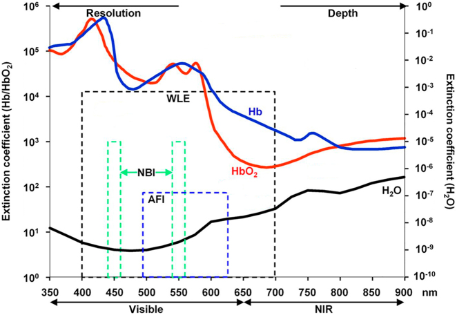
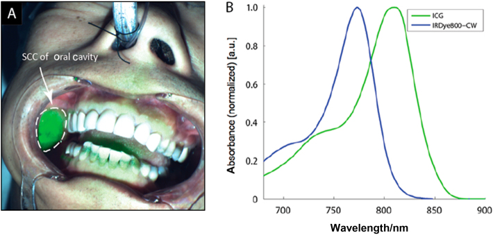
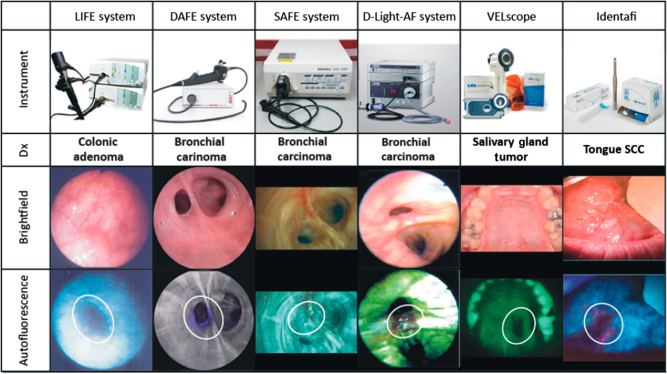
Head and neck cancers become a severe threat to human's health nowadays and represent the sixth most common cancer worldwide. Surgery remains the first-line choice for head and neck cancer patients. Limited resectable tissue mass and complicated anatomy structures in the head and neck region put the surgeons in a dilemma between the extensive resection and a better quality of life for the patients. Early diagnosis and treatment of the pre-malignancies, as well as real-time in vivo detection of surgical margins during en bloc resection, could be leveraged to minimize the resection of normal tissues. With the understanding of the head and neck oncology, recent advances in optical hardware and reagents have provided unique opportunities for real-time pre-malignancies and cancer imaging in the clinic or operating room. Optical imaging in the head and neck has been reported using autofluorescence imaging, targeted fluorescence imaging, high-resolution microendoscopy, narrow band imaging and the Raman spectroscopy. In this study, we reviewed the basic theories and clinical applications of optical imaging for the diagnosis and treatment in the field of head and neck oncology with the goal of identifying limitations and facilitating future advancements in the field.
如今,头颈部癌症对人类健康构成了严重威胁,是全球第六大常见癌症。手术仍然是头颈部癌症患者的首选治疗方法。头颈部区域可切除组织有限且解剖结构复杂,这使得外科医生在广泛切除和提高患者生活质量之间陷入两难境地。早期诊断和治疗癌前病变,以及在整块切除过程中实时活体检测手术切缘,都可以尽量减少正常组织的切除。随着对头颈部肿瘤学的认识不断深入,光学硬件和试剂的最新进展为临床或手术室中实时癌前病变和癌症成像提供了独特的机会。已经有报道使用自发荧光成像、靶向荧光成像、高分辨率显微内镜、窄带成像和拉曼光谱等方法对头颈部进行光学成像。本研究综述了光学成像在头颈部肿瘤学诊断和治疗领域的基础理论和临床应用,旨在确定该领域的局限性并促进未来的发展。