Cheung Yun-Chung, Juan Yu-Hsiang, Lo Yung-Feng, Lin Yu-Ching, Yeh Chih-Hua, Ueng Shir-Hwa
Department of Medical Imaging and Intervention.
Department of Medical Imaging and Radiological Sciences, Medical College of Chang Gung University.
Medicine (Baltimore). 2020 Jan;99(5):e19024. doi: 10.1097/MD.0000000000019024.
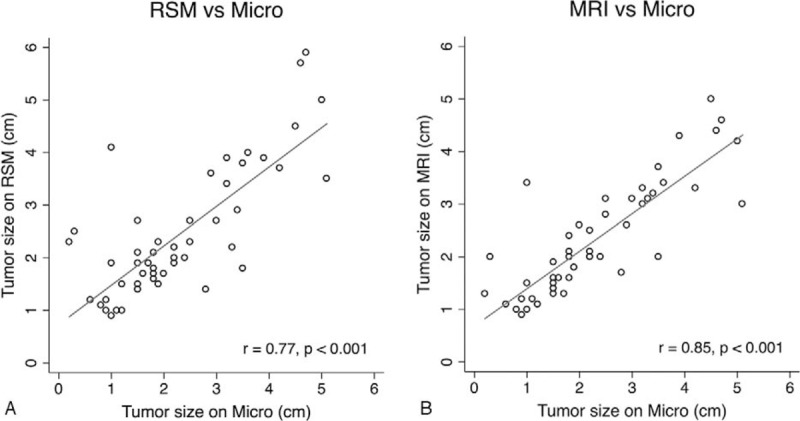
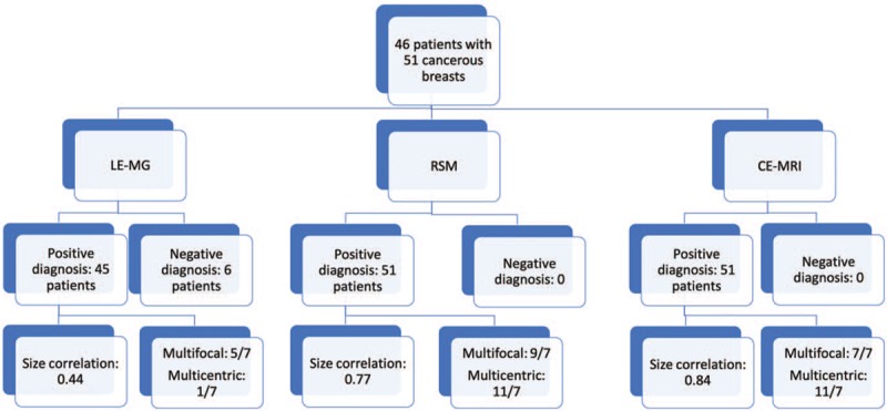
To assess the feasibility of using contrast-enhanced spectral mammography (CESM) for operative planning of patients with breast cancers who were initially diagnosed by sonographic guided biopsy.With the approval of the Institutional Review Board of our hospital, we retrospectively reviewed the data on patients with breast cancers who underwent CESM and contrast-enhanced magnetic resonance imaging (CE-MRI) prior to operation and were followed up for at least 5 years postoperatively. The patients with breast cancer diagnosed by sonographic guided biopsy without mammography were included for analysis. The size and number of cancers on low-energy mammograms (LE-MG), recombined subtracted mammograms (RSM), and CE-MRI were recorded and compared with microscopic histopathologic data and at least 5 years of clinical follow-up data.Fifty-one cancerous breasts of 46 patients were included in the analysis. All the principal cancers could be detected by RSM or CE-MRI; however, only 45 were by LE-MG. The Pearson correlation coefficients for the size on microscopy were 0.44 for LE-MG, 0.77 for RSM, and 0.84 for CE-MRI (all P-values ≤.001). Regarding the microscopic reports, RSM or CE-MRI had sensitivities of 100% and a positive predictive value of 63.6% for multicentric cancers. One breast cancer with partial mastectomy recurred after 3 years of follow-up.CESM was feasible for assessing the cancer extension and multicentric cancers as secondary examination in patients with diagnosed breast cancers after sonographic biopsy.
评估采用对比增强光谱乳腺摄影(CESM)对最初经超声引导活检诊断的乳腺癌患者进行手术规划的可行性。经我院机构审查委员会批准,我们回顾性分析了术前接受CESM和对比增强磁共振成像(CE-MRI)检查且术后至少随访5年的乳腺癌患者的数据。纳入经超声引导活检诊断但未行乳腺摄影的乳腺癌患者进行分析。记录低能量乳腺摄影(LE-MG)、重组减影乳腺摄影(RSM)和CE-MRI上癌灶的大小和数量,并与显微镜下组织病理学数据及至少5年的临床随访数据进行比较。46例患者的51个患癌乳房纳入分析。所有主要癌灶均可通过RSM或CE-MRI检测到;然而,LE-MG仅检测到45个。显微镜下测量大小的Pearson相关系数,LE-MG为0.44,RSM为0.77,CE-MRI为0.84(所有P值≤0.001)。关于显微镜检查报告,RSM或CE-MRI对多中心癌的敏感性为100%,阳性预测值为63.6%。1例接受保乳手术的乳腺癌患者随访3年后复发。CESM作为超声活检后确诊乳腺癌患者的二次检查手段,对于评估癌灶范围及多中心癌是可行的。