Gelardi Fabrizia, Ragaini Elisa Maria, Sollini Martina, Bernardi Daniela, Chiti Arturo
Department of Biomedical Sciences, Humanitas University, Via Rita Levi Montalcini 4, 20072 Pieve Emanuele, Italy.
IRCCS Humanitas Research Hospital, Via Manzoni 56, 20089 Rozzano, Italy.
Diagnostics (Basel). 2022 Aug 4;12(8):1890. doi: 10.3390/diagnostics12081890.
Contrast-enhanced mammography (CEM) and contrast-enhanced magnetic resonance imaging (CE-MRI) are commonly used in the screening of breast cancer. The present systematic review aimed to summarize, critically analyse, and meta-analyse the available evidence regarding the role of CE-MRI and CEM in the early detection, diagnosis, and preoperative assessment of breast cancer.
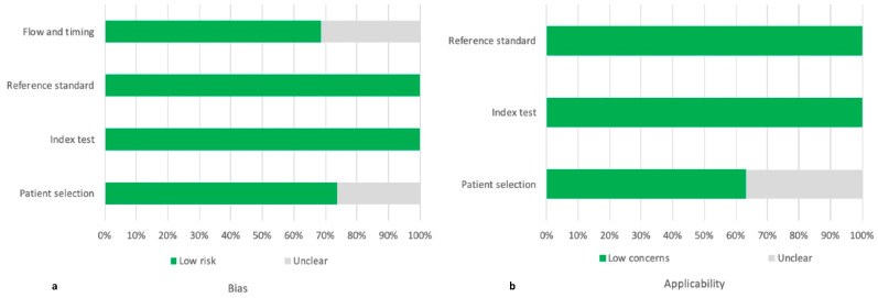
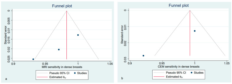
The search was performed on PubMed, Google Scholar, and Web of Science on 28 July 2021 using the following terms "breast cancer", "preoperative staging", "contrast-enhanced mammography", "contrast-enhanced spectral mammography", "contrast enhanced digital mammography", "contrast-enhanced breast magnetic resonance imaging" "CEM", "CESM", "CEDM", and "CE-MRI". We selected only those papers comparing the clinical efficacy of CEM and CE-MRI. The study quality was assessed using the QUADAS-2 criteria. The pooled sensitivities and specificity of CEM and CE-MRI were computed using a random-effects model directly from the STATA "metaprop" command. The between-study statistical heterogeneity was tested (I-statistics).
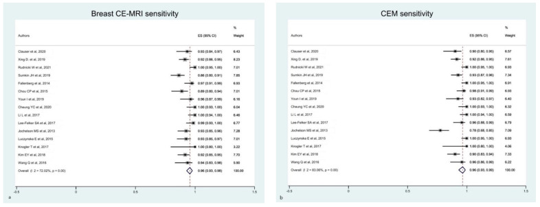
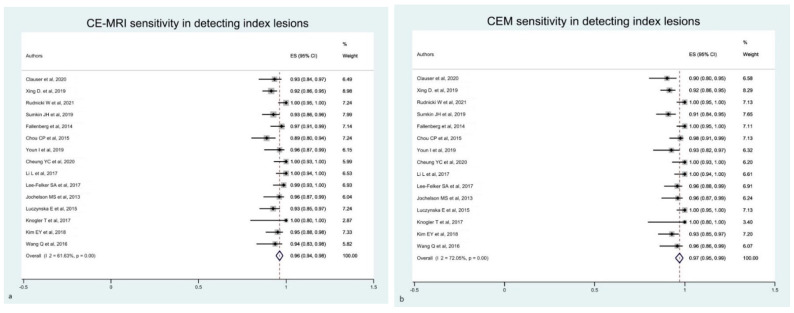
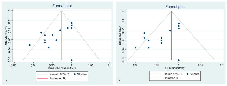
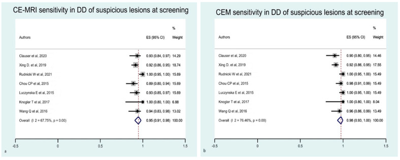
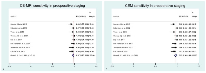
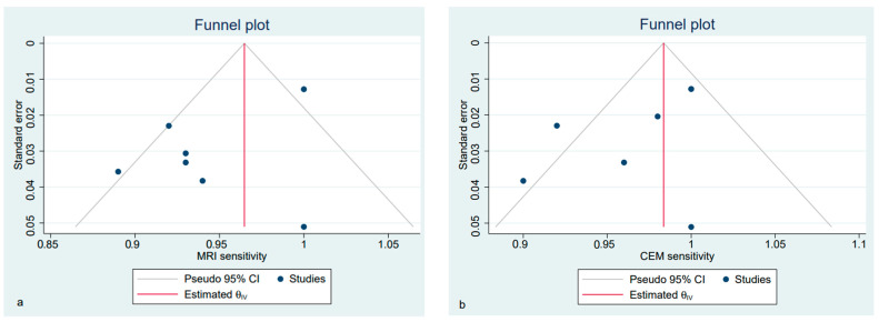
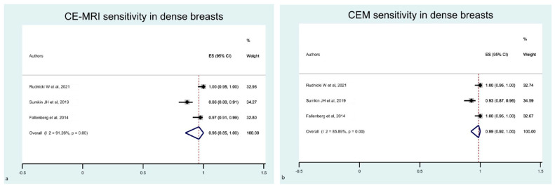
Nineteen studies were selected for this systematic review. Fifteen studies (1315 patients) were included in the metanalysis. Both CEM and CE-MRI detect breast lesions with a high sensitivity, without a significant difference in performance (97% and 96%, respectively).
Our findings confirm the potential of CEM as a supplemental screening imaging modality, even for intermediate-risk women, including females with dense breasts and a history of breast cancer.
对比增强乳腺X线摄影(CEM)和对比增强磁共振成像(CE-MRI)常用于乳腺癌筛查。本系统评价旨在总结、批判性分析和荟萃分析关于CE-MRI和CEM在乳腺癌早期检测、诊断及术前评估中作用的现有证据。
2021年7月28日在PubMed、谷歌学术和科学网进行检索,检索词如下:“乳腺癌”“术前分期”“对比增强乳腺X线摄影”“对比增强光谱乳腺X线摄影”“对比增强数字乳腺X线摄影”“对比增强乳腺磁共振成像”“CEM”“CESM”“CEDM”及“CE-MRI”。我们仅选择那些比较CEM和CE-MRI临床疗效的论文。采用QUADAS-2标准评估研究质量。使用随机效应模型直接从STATA“metaprop”命令计算CEM和CE-MRI的合并敏感度和特异度。检验研究间的统计学异质性(I统计量)。
本系统评价纳入19项研究。荟萃分析纳入15项研究(1315例患者)。CEM和CE-MRI均能以高敏感度检测乳腺病变,性能无显著差异(分别为97%和96%)。
我们的研究结果证实了CEM作为一种补充筛查成像方式的潜力,即使对于中度风险女性,包括乳腺致密和有乳腺癌病史的女性也是如此。