Partain Brittany D, Unni Mythreyi, Rinaldi Carlos, Allen Kyle D
J. Crayton Pruitt Family Department of Biomedical Engineering, University of Florida, Gainesville, FL, USA.
Department of Chemical Engineering, University of Florida, Gainesville, FL, USA.
J Control Release. 2020 May 10;321:259-271. doi: 10.1016/j.jconrel.2020.01.052. Epub 2020 Jan 28.
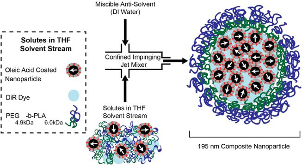
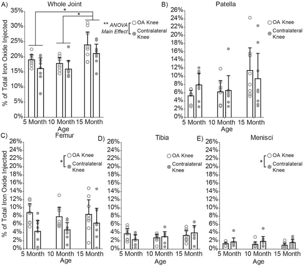
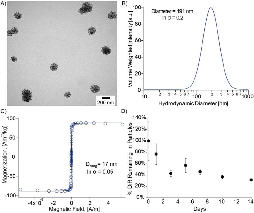
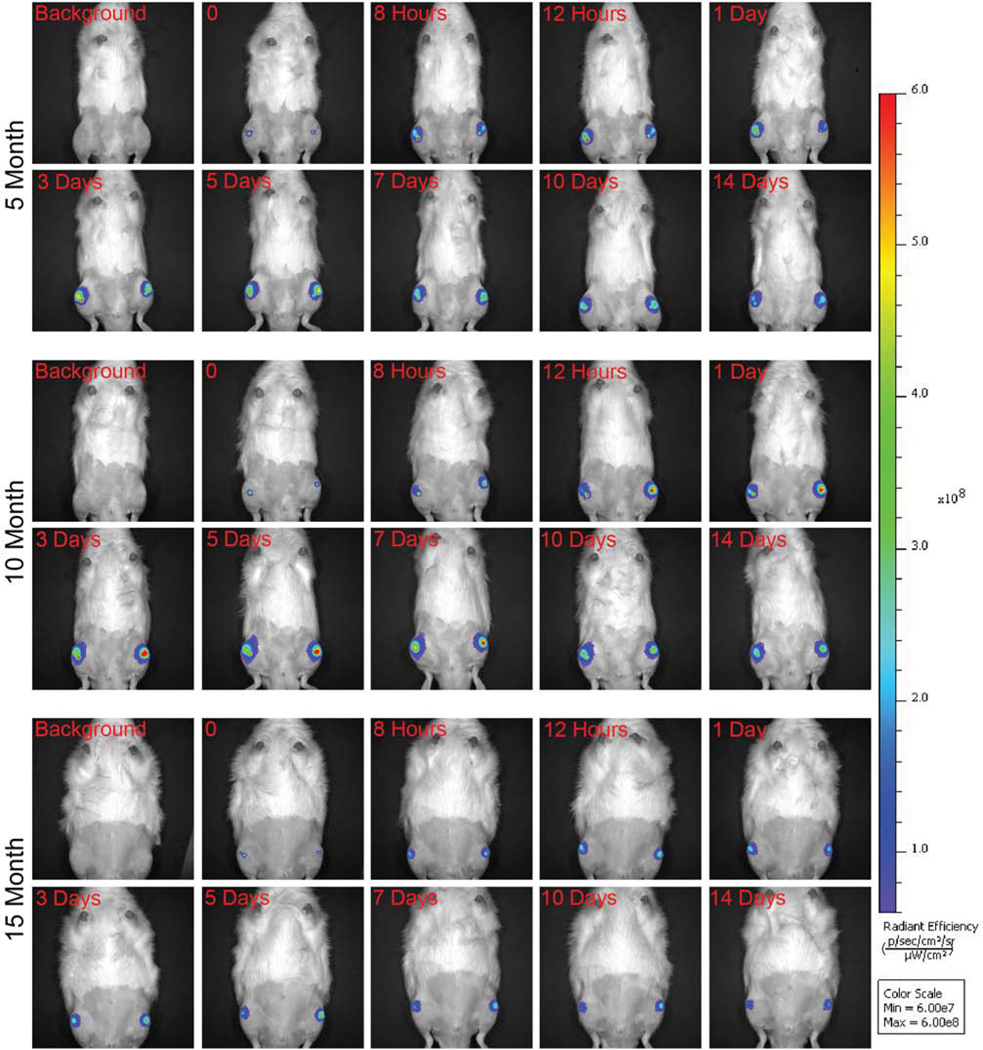
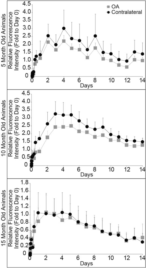
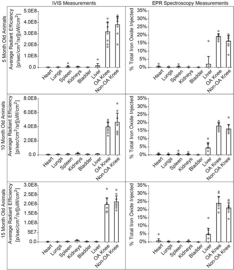
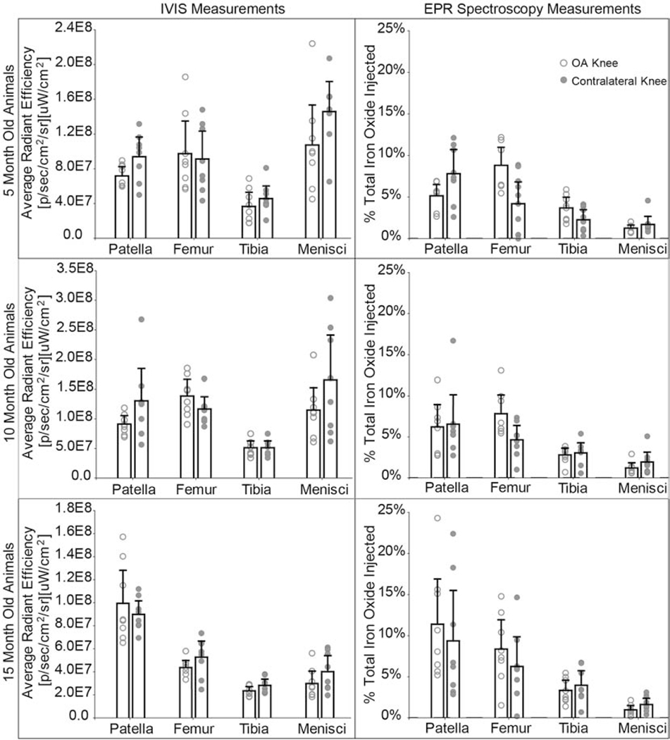
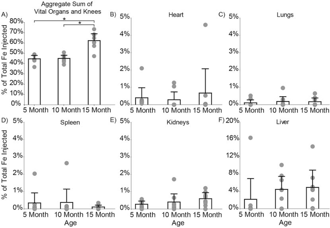
Intra-articular injections are the most direct route for administering osteoarthritis (OA) therapies, yet how drug carriers distribute within the joint remains understudied. To this end, we developed a magnetic composite nanoparticle that can be tracked with fluorescence in vivo via an in vivo imaging system (IVIS), and quantified ex vivo via electron paramagnetic resonance (EPR) spectroscopy. Using this particle, the effects of age and OA pathogenesis on particle clearance and distribution were evaluated in the medial meniscus transection model of OA (5-, 10-, and 15-month old male Lewis rats). At 9 weeks after meniscus transection, composite nanoparticles were injected and joint clearance was assessed via IVIS. At 2 weeks after injection, animals were euthanized and particle distribution was quantified ex vivo via EPR spectroscopy. IVIS and EPR spectroscopy data indicate a predominant amount of particles remained in the joint after 14 days. EPR spectroscopy data suggests particles cleared more slowly from OA knees than from the contralateral control, with particles clearing more slowly from 15-month old rats than from 5- and 10-month old rats. This study demonstrates the importance of including both age and OA as factors when evaluating nanoparticles for intra-articular drug delivery.
关节内注射是给予骨关节炎(OA)治疗药物最直接的途径,但药物载体在关节内的分布情况仍未得到充分研究。为此,我们开发了一种磁性复合纳米颗粒,它可以通过体内成像系统(IVIS)在体内用荧光进行追踪,并通过电子顺磁共振(EPR)光谱在体外进行定量分析。利用这种颗粒,在OA的内侧半月板横断模型(5、10和15月龄雄性Lewis大鼠)中评估了年龄和OA发病机制对颗粒清除和分布的影响。在半月板横断9周后,注射复合纳米颗粒,并通过IVIS评估关节清除情况。注射2周后,对动物实施安乐死,并通过EPR光谱在体外对颗粒分布进行定量分析。IVIS和EPR光谱数据表明,14天后大部分颗粒仍留在关节内。EPR光谱数据表明,颗粒从OA膝关节清除的速度比从对侧对照膝关节慢,且从15月龄大鼠体内清除的速度比从5月龄和10月龄大鼠体内慢。这项研究证明了在评估用于关节内给药的纳米颗粒时,将年龄和OA都作为因素考虑的重要性。