Department of Clinical Science, Umeå University, Umeå, Sweden.
Wallenberg Center for Molecular Medicine (WCMM), Umeå University, Umeå, Sweden.
Eur Radiol. 2020 May;30(5):2543-2551. doi: 10.1007/s00330-019-06636-4. Epub 2020 Jan 31.
Assess the sensitivity and specificity of computed tomography angiography (CTA) for carotid near-occlusion diagnosis interpreted in clinical practice against expert assessment.
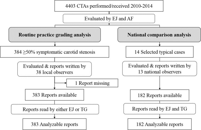
CTAs were graded by two expert interpreters for near-occlusion. Findings were compared with clinical reports in 383 consecutive cases with symptomatic ≥ 50% carotid stenosis. In addition, 14 selected CTA exams (8 near-occlusions and 6 controls) were analyzed in a national effort by 13 radiologists experienced with carotid CTA.
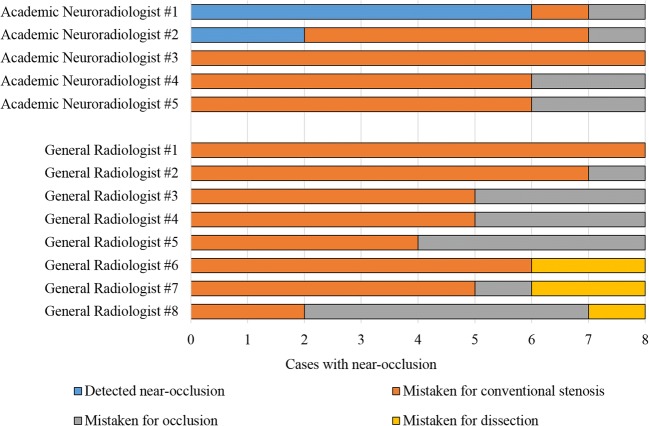
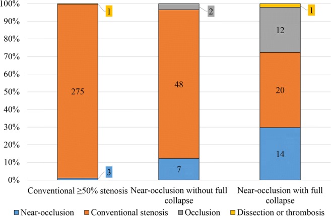
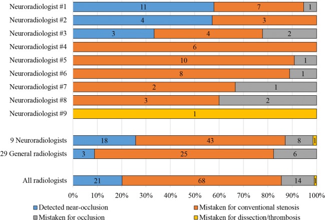
In clinical practice, imaging reports were 20% (95% CI 12-28%) sensitive for near-occlusion, ranging 0-58% between different radiologists; specificity was 99%. Among the 13 radiologists reviewing the same 8 near-occlusions, the average sensitivity was 8%, ranging 0-75%; specificity was 100%.
Carotid near-occlusion is systematically under-reported in clinical routine practice, caused by limited application of grading criteria when assessing CTA.
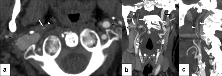
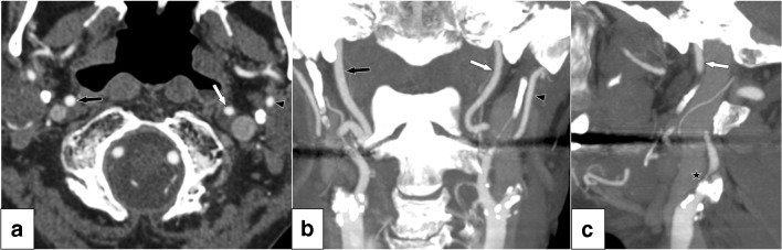
• Carotid near-occlusion is severe stenosis with distal artery collapse; this collapse is often subtle. • A fifth of near-occlusions were detected in routine practice. Many readers mistake near-occlusion for stenosis without distal artery collapse, either by not actively searching for subtle collapses or by not interpreting the collapse correctly when noticed. • On the other hand, the novice diagnostician should be cautioned to not over-diagnose near-occlusion; other causes of extracranial ICA asymmetry also exist such as distal disease and Circle of Willis anatomical variants.
评估在临床实践中解读的计算机断层血管造影(CTA)对颈动脉近闭塞诊断的敏感性和特异性,与专家评估相比。
由两位专家解读者对 CTA 进行近闭塞分级。在 383 例有症状≥50%颈动脉狭窄的连续病例中,将检查结果与临床报告进行比较。此外,通过 13 位具有颈动脉 CTA 经验的放射科医生对 14 例选定的 CTA 检查(8 例近闭塞和 6 例对照)进行了全国性分析。
在临床实践中,影像学报告对近闭塞的敏感性为 20%(95%可信区间为 12-28%),不同放射科医生之间的范围为 0-58%;特异性为 99%。在对 8 例近闭塞进行相同评估的 13 位放射科医生中,平均敏感性为 8%,范围为 0-75%;特异性为 100%。
在临床常规实践中,颈动脉近闭塞被系统地漏报,这是由于在评估 CTA 时对分级标准的应用有限。
颈动脉近闭塞是严重狭窄伴远端动脉塌陷;这种塌陷通常很细微。
在常规实践中发现近闭塞的比例为五分之一。许多读者错误地将近闭塞诊断为没有远端动脉塌陷的狭窄,要么是因为没有积极地寻找细微的塌陷,要么是因为当注意到塌陷时没有正确地解释它。
另一方面,初学者应注意不要过度诊断近闭塞;颅外颈内动脉不对称的其他原因也存在,如远端疾病和 Willis 环解剖变异。