Shaikh Kashif, Kinninger April, Cherukuri Lavanya, Birudaraju Divya, Nakanishi Rine, Almeida Shone, Jayawardena Eranthi, Shekar Chandana, Flores Ferdinand, Hamal Sajad, Sheikh Mohammed Salman, Johanis Amit, Cu Benedict, Budoff Matthew J
Lundquist Institute for Biomedical Innovation at Harbor UCLA Medical Center, Torrance, CA 90502, USA.
Exp Ther Med. 2020 Feb;19(2):1457-1461. doi: 10.3892/etm.2019.8371. Epub 2019 Dec 27.
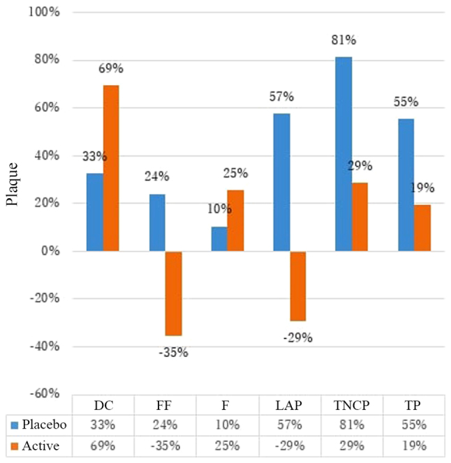
Several previous studies have demonstrated that aged garlic extract (AGE) inhibits the progression of coronary artery calcification and non-calcified plaque (NCP) in the general population. However, its effects on plaque progression in patients with diabetes have not yet been investigated, at least to the best of our knowledge. This study investigated whether AGE reduces the coronary plaque volume measured by cardiac computed tomography angiography (CCTA) in patients with diabetes mellitus (DM). A total of 80 participants with DM with a median age of 57 years were prospectively assigned to consume 2,400 mg AGE/day (after completion, 37 participants) or placebo (after completion, 29 participants) orally. Both groups underwent CCTA at baseline and follow-up 365 days apart. In total, 66 participants completed the study. Coronary plaque volume, including total plaque (TP), dense calcium (DC), fibrous, fibro-fatty and low-attenuation plaque (LAP) volumes were measured based upon pre-defined intensity cut-off values using semi-automated software (QAngio CT). Changes in various plaque types were normalized to the total coronary artery length. The non-parametric Wilcoxon rank-sum test was performed to examine the differences in plaque formation between the 2 groups. No significant differences were found in the baseline characteristics between the AGE and placebo groups. Compared with the placebo group, the AGE group exhibited a statistically significant regression in normalized LAP [median and standard deviation (SD) -0.2 (18.8) vs. 2.5 (69.3), P=0.0415]. No differences were observed in TP, fibrous, or fibrofatty plaque volumes between the AGE and placebo group. On the whole, this study indicated that the %LAP change in the AGE group was significantly greater than that in the placebo group in patients with diabetes. However, further studies are warranted to evaluate whether AGE has the ability to stabilize vulnerable plaque and decrease adverse cardiovascular events.
此前的多项研究表明,在普通人群中, aged garlic extract (AGE) 可抑制冠状动脉钙化和非钙化斑块(NCP)的进展。然而,据我们所知,其对糖尿病患者斑块进展的影响尚未得到研究。本研究调查了AGE是否能减少糖尿病(DM)患者通过心脏计算机断层扫描血管造影(CCTA)测量的冠状动脉斑块体积。共有80名年龄中位数为57岁的DM参与者被前瞻性地分配为口服2400 mg AGE/天(完成后37名参与者)或安慰剂(完成后29名参与者)。两组在基线时和间隔365天的随访时均接受了CCTA检查。共有66名参与者完成了研究。基于预先定义的强度截止值,使用半自动软件(QAngio CT)测量冠状动脉斑块体积,包括总斑块(TP)、致密钙(DC)、纤维、纤维脂肪和低衰减斑块(LAP)体积。将各种斑块类型的变化标准化为冠状动脉总长度。采用非参数Wilcoxon秩和检验来检验两组之间斑块形成的差异。AGE组和安慰剂组的基线特征没有显著差异。与安慰剂组相比,AGE组在标准化LAP方面表现出统计学上的显著消退[中位数和标准差(SD)为-0.2(18.8)对2.5(69.3),P=0.0415]。AGE组和安慰剂组在TP、纤维或纤维脂肪斑块体积方面没有观察到差异。总体而言,本研究表明,糖尿病患者中AGE组的%LAP变化显著大于安慰剂组。然而,需要进一步研究来评估AGE是否有能力稳定易损斑块并减少不良心血管事件。