Department of Biochemistry, Indian Institute of Science, Bangalore, India.
Indira Clinic, Bangalore, India.
Front Cell Infect Microbiol. 2020 Jan 14;9:430. doi: 10.3389/fcimb.2019.00430. eCollection 2019.
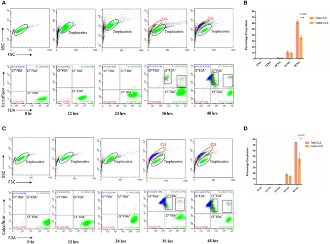
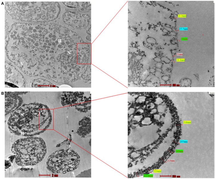
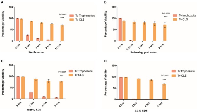
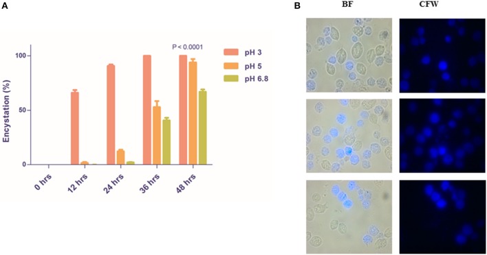
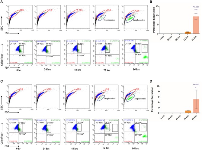
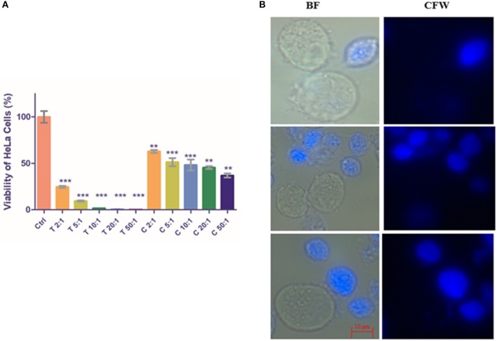
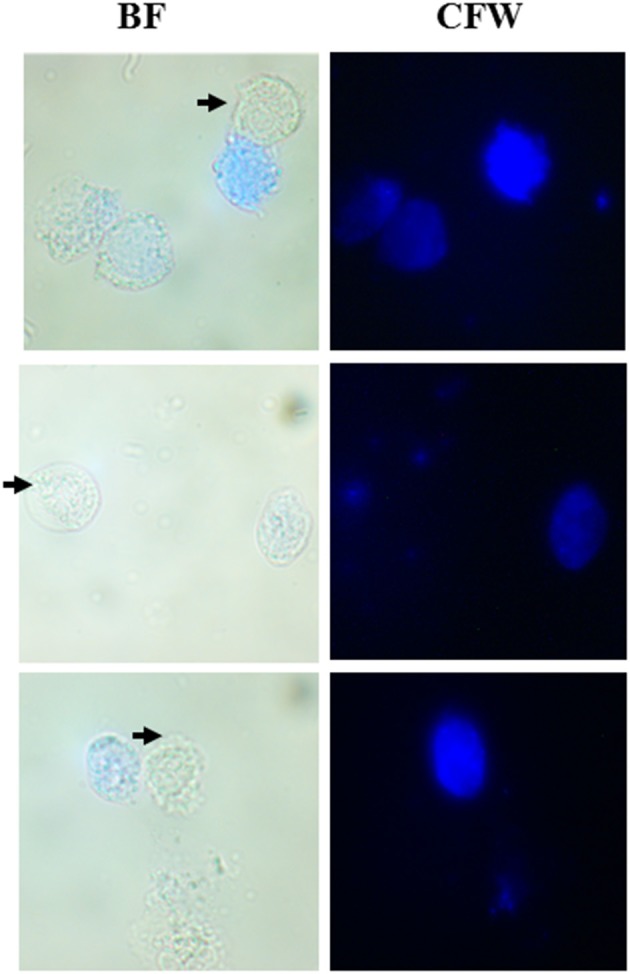
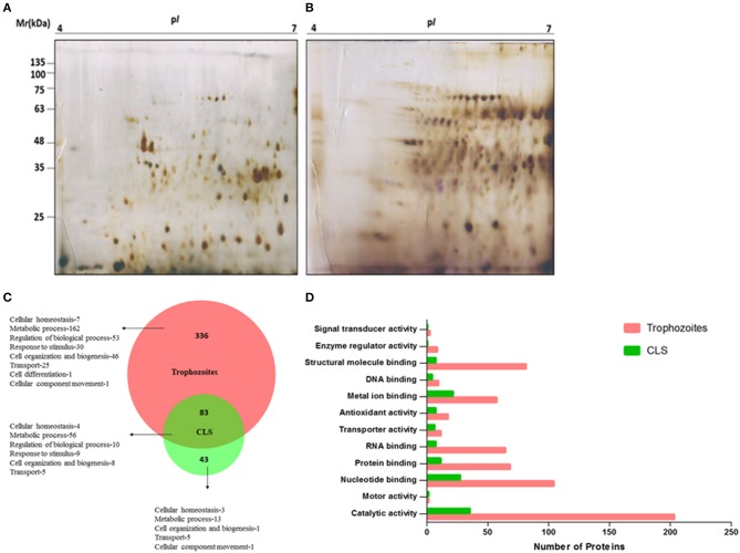
is the parasitic protozoan residing in human urogenital tract causing trichomoniasis, which is the leading non-viral sexually transmitted disease. It has cosmopolitan distribution throughout the globe and affects both men and women. Lifecycle of the parasite has been traditionally described as consisting of motile and symptom-causing trophozoites. Chemical and temperature perturbations in trophozoites have been shown to aid conversion to pseudocysts, which is poorly investigated. In the current study, we show the formation of viable cyst-like structures (CLS) in stationary phase of axenic culture. We used a fluorescent stain called calcofluor white, which specifically binds to chitin and cellulose-containing structures, to score for CLS. Using flow cytometry, we demonstrated and quantitated the processes of encystation as well as excystation; thus, completing the parasite's lifecycle without any chemical/temperature alterations. Like cysts from other protozoan parasites such as and CLS appeared spherical, immotile, and resistant to osmotic lysis and detergent treatments. Ultrastructure of CLS demonstrated by Transmission Electron Microscopy showed a thick electron-dense deposition along its outer membrane. To probe the physiological role of CLS, we exposed parasites to vaginal pH and observed that trophozoites took this as a cue to convert to CLS. Further, upon co- culturing with cells of cervical origin, CLS rapidly excysted to form trophozoites which abrogated the cervical cell monolayer in a dose-dependent manner. To further corroborate the presence of two distinct forms in , we performed two-dimensional gel electrophoresis and global, untargeted mass spectrometry to highlight differences in the proteome with trophozoites. Interestingly, CLS remained viable in chlorinated swimming pool water implicating the possibility of its role as environmentally resistant structures involved in non-sexual mode of parasite transmission. Finally, we showed that symptomatic human patient vaginal swabs had both trophozoites and CLS; thus, highlighting its importance in clinical infections. Overall, our study highlights the plasticity of the pathogen and its rapid adaption when subjected to stressful environmental cues and suggests an important role of CLS in the parasite's life cycle, pathogenesis and transmission.
是一种寄生的原生动物,寄生于人体泌尿生殖道,引起滴虫病,是主要的非病毒性性传播疾病。它在全球范围内广泛分布,影响男性和女性。寄生虫的生命周期传统上被描述为由运动和引起症状的滋养体组成。已经表明,滋养体中的化学和温度扰动有助于转化为伪囊肿,但对其研究甚少。在本研究中,我们在无菌培养的静止期观察到了可行的囊样结构(CLS)的形成。我们使用了一种称为钙荧光白的荧光染料,它专门与含有几丁质和纤维素的结构结合,以对 CLS 进行评分。使用流式细胞术,我们证明并量化了包囊形成和脱囊过程;因此,在没有任何化学/温度改变的情况下完成了寄生虫的生命周期。与其他原生动物寄生虫(如 和 )的囊肿一样,CLS 呈球形,无运动性,对渗透压裂解和去污剂处理具有抗性。透射电子显微镜显示的 CLS 的超微结构表明,其外膜上有一层厚厚的电子致密沉积物。为了探究 CLS 的生理作用,我们将寄生虫暴露于阴道 pH 值下,观察到滋养体将其作为转化为 CLS 的信号。此外,当与宫颈来源的细胞共培养时,CLS 迅速脱囊形成滋养体,以剂量依赖的方式破坏宫颈细胞单层。为了进一步证实 中存在两种不同的形式,我们进行了二维凝胶电泳和全局非靶向质谱分析,以突出滋养体与蛋白质组的差异。有趣的是,CLS 在氯化游泳池水中仍然存活,这暗示了其作为参与非性传播寄生虫的环境抗性结构的可能性。最后,我们表明,有症状的人类患者阴道拭子既有滋养体又有 CLS;因此,强调了其在临床感染中的重要性。总的来说,我们的研究强调了病原体的可塑性及其在受到应激环境线索时的快速适应,并表明 CLS 在寄生虫生命周期、发病机制和传播中具有重要作用。