Department of Endocrinology, The Affiliated Lianyungang Hospital of Xuzhou Medical University, The First People's Hospital of Lianyungang, Lianyungang Clinical College of Nanjing Medical University, Lianyungang 222000, China.
Department of Gastrointestinal Surgery, The Affiliated Lianyungang Hospital of Xuzhou Medical University, The First People's Hospital of Lianyungang, Lianyungang Clinical College of Nanjing Medical University, Lianyungang 222000, China.
Biosci Rep. 2020 Mar 27;40(3). doi: 10.1042/BSR20193800.
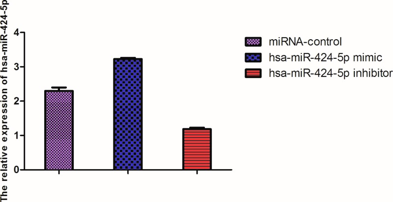
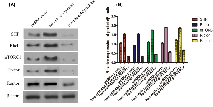
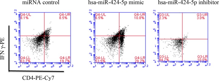
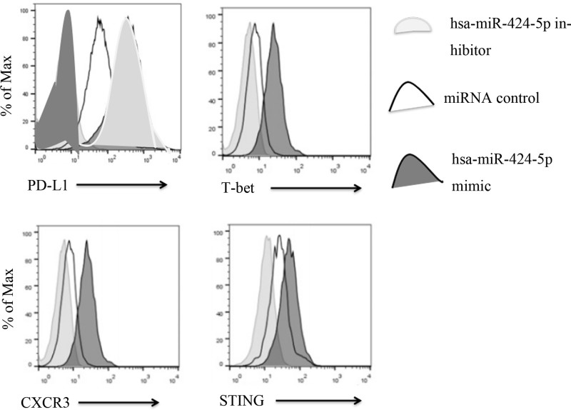
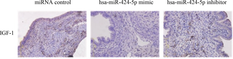
In the present study, hsa-miR-424-5p mimic plasmid and hsa-mir-424-5p inhibitor plasmid were designed and injected into rats respectively, and miRNA control plasmid was also constructed. Models of Type 1 diabetes (T1D) were built. After successful modeling, the expression of hsa-miR-424-5p in lymphocytes was analyzed by RT-PCR. The expression of protein PD-1, T-bet, CXCR3, STING in Th1 lymphocytes and content of IGF-1 in islet tissue were analyzed by flow analysis. The protein levels of SHP2, Rheb, mTORC1, Rictor and Raptor in islet tissue were analyzed by Western blot. The results showed that hsa-miR-424-5p mimic group had the highest expression of hsa-miR-424-5p in lymphocytes. The expression of PD-1 was in hsa-miR-424-5p inhibitor > miRNA control > hsa-miR-424-5p mimic, while the expression of T-bet, CXCR3 and STING was in hsa-miR-424-5p mimic > miRNA control > hsa-miR-424-5p inhibitor. The expression of IGF-1 protein in hsa-miR-424-5p inhibitor group was the highest (32.08%) and hardly expressed in hsa-miR-424-5p mimic group (2.36%). The expression of SHP2, Rheb, mTORC1, Rictor and Raptor of insulin histoproteins were in hsa-miR-424-5p mimic group > miRNA control of > hsa-miR-424-5p inhibitor group, with statistical differences. It indicates that hsa-miR-424-5p binding PD-1 signaling molecules can stimulate the immune effect through the mTORC signaling pathway and participates in the pathogenesis of T1D.
在本研究中,分别设计了 hsa-miR-424-5p 模拟质粒和 hsa-mir-424-5p 抑制剂质粒,并构建了 miRNA 对照质粒。构建 1 型糖尿病(T1D)模型。建模成功后,通过 RT-PCR 分析淋巴细胞中 hsa-miR-424-5p 的表达。通过流式分析分析 Th1 淋巴细胞中 PD-1、T-bet、CXCR3、STING 的表达以及胰岛组织中 IGF-1 的含量。通过 Western blot 分析胰岛组织中 SHP2、Rheb、mTORC1、Rictor 和 Raptor 的蛋白水平。结果表明,hsa-miR-424-5p 模拟组淋巴细胞中 hsa-miR-424-5p 表达最高。PD-1 的表达为 hsa-miR-424-5p 抑制剂>miRNA 对照>hsa-miR-424-5p 模拟,而 T-bet、CXCR3 和 STING 的表达为 hsa-miR-424-5p 模拟>miRNA 对照>hsa-miR-424-5p 抑制剂。hsa-miR-424-5p 抑制剂组 IGF-1 蛋白表达最高(32.08%),hsa-miR-424-5p 模拟组几乎不表达(2.36%)。胰岛素组织蛋白 SHP2、Rheb、mTORC1、Rictor 和 Raptor 的表达为 hsa-miR-424-5p 模拟组>miRNA 对照组>hsa-miR-424-5p 抑制剂组,差异有统计学意义。表明 hsa-miR-424-5p 结合 PD-1 信号分子,通过 mTORC 信号通路刺激免疫效应,参与 T1D 的发病机制。