Department of Neurosurgery, The First Hospital of Jilin University, No. 1 Xinmin Avenue, Changchun, 130021, Jilin, People's Republic of China.
Department of Neurology, The First Hospital of Jilin University, Changchun, 130021, People's Republic of China.
J Neuroinflammation. 2020 Feb 3;17(1):46. doi: 10.1186/s12974-020-1725-8.
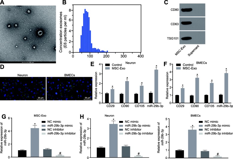
Mesenchymal stem cells (MSCs) are suspected to exert neuroprotective effects in brain injury, in part through the secretion of extracellular vesicles like exosomes containing bioactive compounds. We now investigate the mechanism by which bone marrow MSCs (BMSCs)-derived exosomes harboring the small non-coding RNA miR-29b-3p protect against hypoxic-ischemic brain injury in rats.
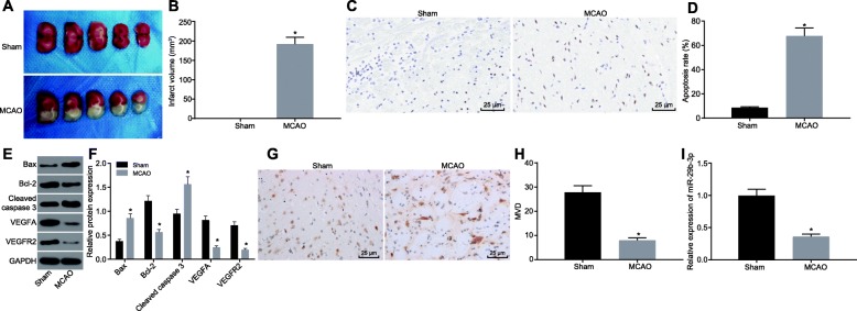
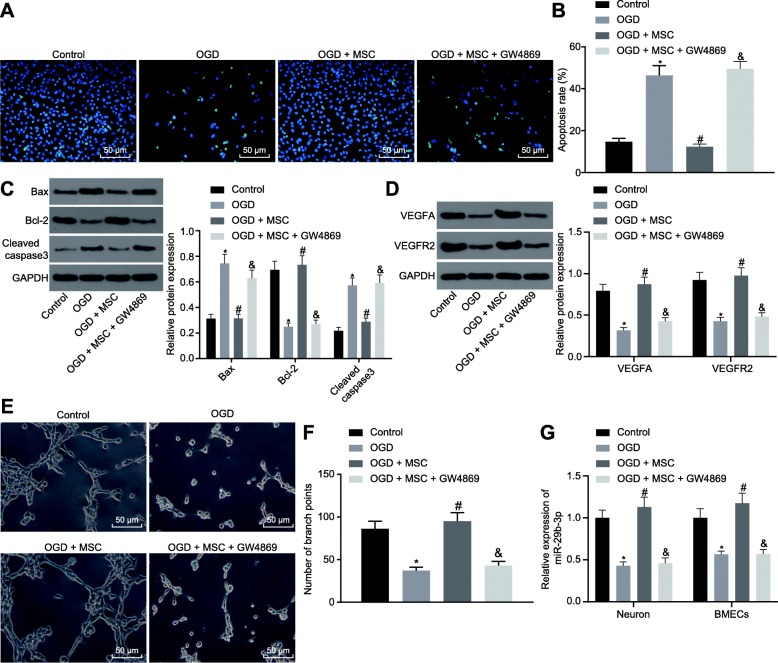
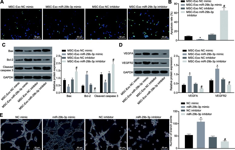
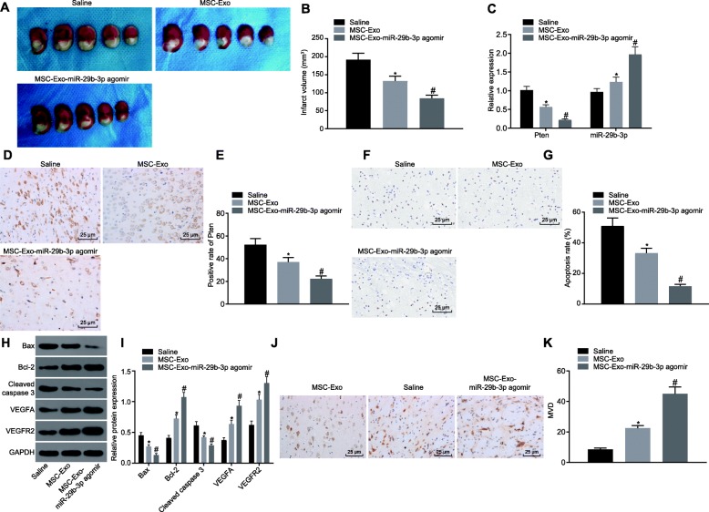
We established a rat model of middle cerebral artery occlusion (MCAO) and primary cortical neuron or brain microvascular endothelial cell (BMEC) models of oxygen and glucose deprivation (OGD). Exosomes were isolated from the culture medium of BMSCs. We treated the MCAO rats with BMSC-derived exosomes in vivo, and likewise the OGD-treated neurons and BMECs in vitro. We then measured apoptosis- and angiogenesis-related features using TUNEL and CD31 immunohistochemical staining and in vitro Matrigel angiogenesis assays.
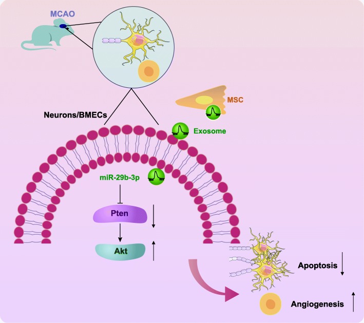
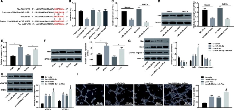
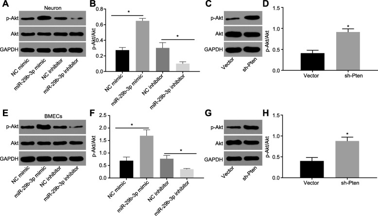
The dual luciferase reporter gene assay showed that miR-29b-3p targeted the protein phosphatase and tensin homolog (PTEN). miR-29b-3p was downregulated and PTEN was upregulated in the brain of MCAO rats and in OGD-treated cultured neurons. MCAO rats and OGD-treated neurons showed promoted apoptosis and decreased angiogenesis, but overexpression of miR-29b-3p or silencing of PTEN could reverse these alterations. Furthermore, miR-29b-3p could negatively regulate PTEN and activate the Akt signaling pathway. BMSCs-derived exosomes also exerted protective effects against apoptosis of OGD neurons and cell apoptosis in the brain samples from MCAO rats, where we also observed promotion of angiogenesis.
BMSC-derived exosomal miR-29b-3p ameliorates ischemic brain injury by promoting angiogenesis and suppressing neuronal apoptosis, a finding which may be of great significance in the treatment of hypoxic-ischemic brain injury.
间充质干细胞(MSCs)被怀疑通过分泌含有生物活性化合物的细胞外囊泡(如外泌体)发挥对脑损伤的神经保护作用。我们现在研究骨髓间充质干细胞(BMSCs)来源的含有小非编码 RNA miR-29b-3p 的外泌体通过何种机制保护大鼠缺氧缺血性脑损伤。
我们建立了大脑中动脉闭塞(MCAO)大鼠模型和原代皮质神经元或脑微血管内皮细胞(BMEC)氧葡萄糖剥夺(OGD)模型。从 BMSCs 培养上清液中分离出外泌体。我们在体内用 BMSC 来源的外泌体处理 MCAO 大鼠,同样在体外用 OGD 处理的神经元和 BMEC 进行处理。然后通过 TUNEL 和 CD31 免疫组化染色和体外 Matrigel 血管生成测定来测量细胞凋亡和血管生成相关特征。
双荧光素酶报告基因检测表明 miR-29b-3p 靶向蛋白磷酸酶和张力蛋白同源物(PTEN)。MCAO 大鼠脑和 OGD 处理的培养神经元中 miR-29b-3p 下调,PTEN 上调。MCAO 大鼠和 OGD 处理的神经元表现出促进的细胞凋亡和减少的血管生成,但 miR-29b-3p 的过表达或 PTEN 的沉默可以逆转这些改变。此外,miR-29b-3p 可以负调控 PTEN 并激活 Akt 信号通路。BMSCs 来源的外泌体还可以防止 OGD 神经元的细胞凋亡和 MCAO 大鼠脑样本中的细胞凋亡,并观察到促进血管生成。
BMSC 来源的外泌体 miR-29b-3p 通过促进血管生成和抑制神经元凋亡来改善缺血性脑损伤,这一发现对于缺氧缺血性脑损伤的治疗可能具有重要意义。