Department of Neurosurgery, Kanazawa University,13-1 Takara-machi, Kanazawa, 920-8641 Japan; Department of Neurosurgery, The First Hospital of Jilin University, China.
Department of Neurosurgery, Kanazawa University,13-1 Takara-machi, Kanazawa, 920-8641 Japan.
Neuroimage Clin. 2020;25:102192. doi: 10.1016/j.nicl.2020.102192. Epub 2020 Jan 22.
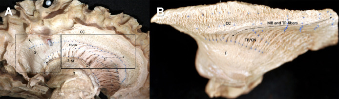
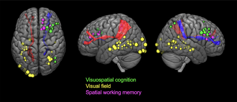
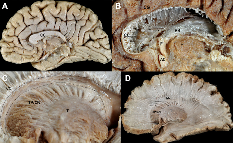
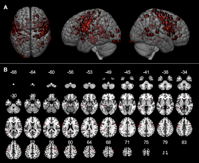
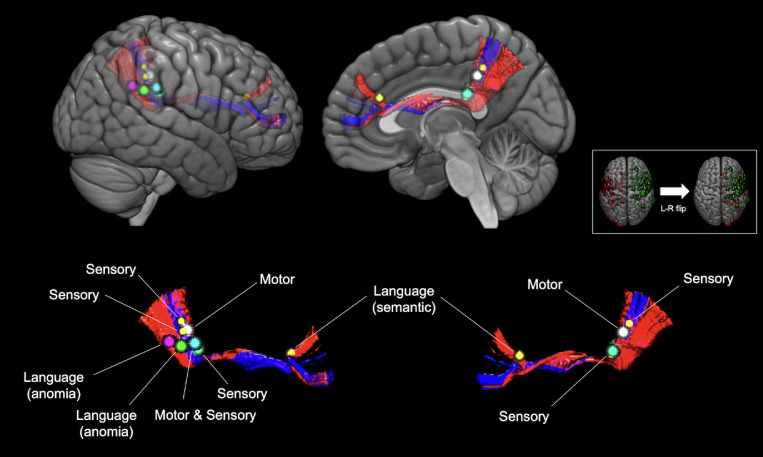
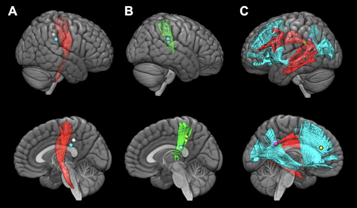
The presence of the superior fronto-occipital fascicle (SFOF) has been reported in the Rhesus monkey; however, it is a subject of controversy in humans. The aim of this study is to identify the SFOF using both in vitro and in vivo anatomo-functional analyses. This study consisted of two approaches. First, one acallosal brain and 12 normal postmortem hemispheres (five left and seven right sides) were dissected under a microscope using Klingler's fiber dissection technique. We focused on the medial subcallosal area superior to the Muratoff bundle, which has been indicated as a principal target area of the SFOF in previous studies. Second, 90 patients underwent awake craniotomy for gliomas with direct electrical stimulations. Functional examinations for visual, ataxic, and cognitive tasks were performed and 453 positive mapping sites were investigated by voxel-based morphometry analysis to establish the functions of the SFOF. The corticostriatal fibers, or the Muratoff bundle, and thalamic peduncle fibers joined in the area of the caudate nucleus, making thalamic peduncle/ corticostriatal bundles, which ran antero-posteriorly in the anterior subcallosal area and radiated from the caudate superior margin in the posterior subcallosal area. However, no SFOF fiber bundle crossed perpendicular to the thalamic peduncle/ corticostriatal bundles in the posterior subcallosal area. In the acallosal hemispheres, Probst bundles were confirmed and the subcallosal areas did not show a specific organization different from the normal brain. Hence, we could not detect a long and continuous association fascicle connecting the frontal lobe and occipital or parietal lobe in the target areas. Furthermore, in the in vivo functional mappings of awake surgery and voxel-based morphometry analysis, eight positive points on the SFOF were selected from the total 453 positive points, but their functions were not related with visual processing and spatial awareness, as has been reported in previous studies. In conclusion, in the present study we attempted to investigate the existence of the SFOF using an anatomical and functional approach. According to our results, the SFOF may not exist in the human brain.
额顶连合束(SFOF)在恒河猴中已有报道,但在人类中存在争议。本研究旨在通过体外和体内解剖功能分析来鉴定 SFOF。本研究包括两个方法。首先,使用 Klingler 的纤维解剖技术,在显微镜下解剖了 1 个无脑联合脑和 12 个正常死后半球(5 个左侧和 7 个右侧)。我们专注于 Muratoff 束上方的内侧亚皮质下区域,这在以前的研究中被认为是 SFOF 的主要靶区。其次,90 名接受清醒开颅手术治疗胶质瘤的患者接受了直接电刺激。进行了视觉、共济失调和认知任务的功能检查,并通过体素形态计量学分析研究了 453 个阳性映射部位,以确定 SFOF 的功能。皮质纹状体纤维,即 Muratoff 束,和丘脑束纤维在尾状核区域结合,形成丘脑束/皮质纹状体束,在前段亚皮质下区域从前向后走行,并从尾状核上缘辐射到后段亚皮质下区域。然而,在后段亚皮质下区域,没有 SFOF 纤维束与丘脑束/皮质纹状体束垂直交叉。在无脑联合半球中,确认了 Probst 束,而亚皮质下区域没有表现出与正常大脑不同的特定组织。因此,我们无法在靶区检测到连接额叶和枕叶或顶叶的长而连续的联合束。此外,在清醒手术的体内功能映射和体素形态计量学分析中,从总共 453 个阳性点中选择了 8 个 SFOF 的阳性点,但它们的功能与视觉处理和空间意识无关,这与以前的研究报道一致。总之,在本研究中,我们试图通过解剖学和功能方法来研究 SFOF 的存在。根据我们的结果,SFOF 可能不存在于人类大脑中。