Center for Morphometric Analysis and Departments of Psychiatry and Neurology, Massachusetts General Hospital, Boston, Massachusetts, USA.
Psychiatry Neuroimaging Laboratory, Department of Psychiatry, Brigham and Women's Hospital, Boston, Massachusetts, USA.
J Comp Neurol. 2023 Dec;531(18):2172-2184. doi: 10.1002/cne.25562. Epub 2023 Nov 27.
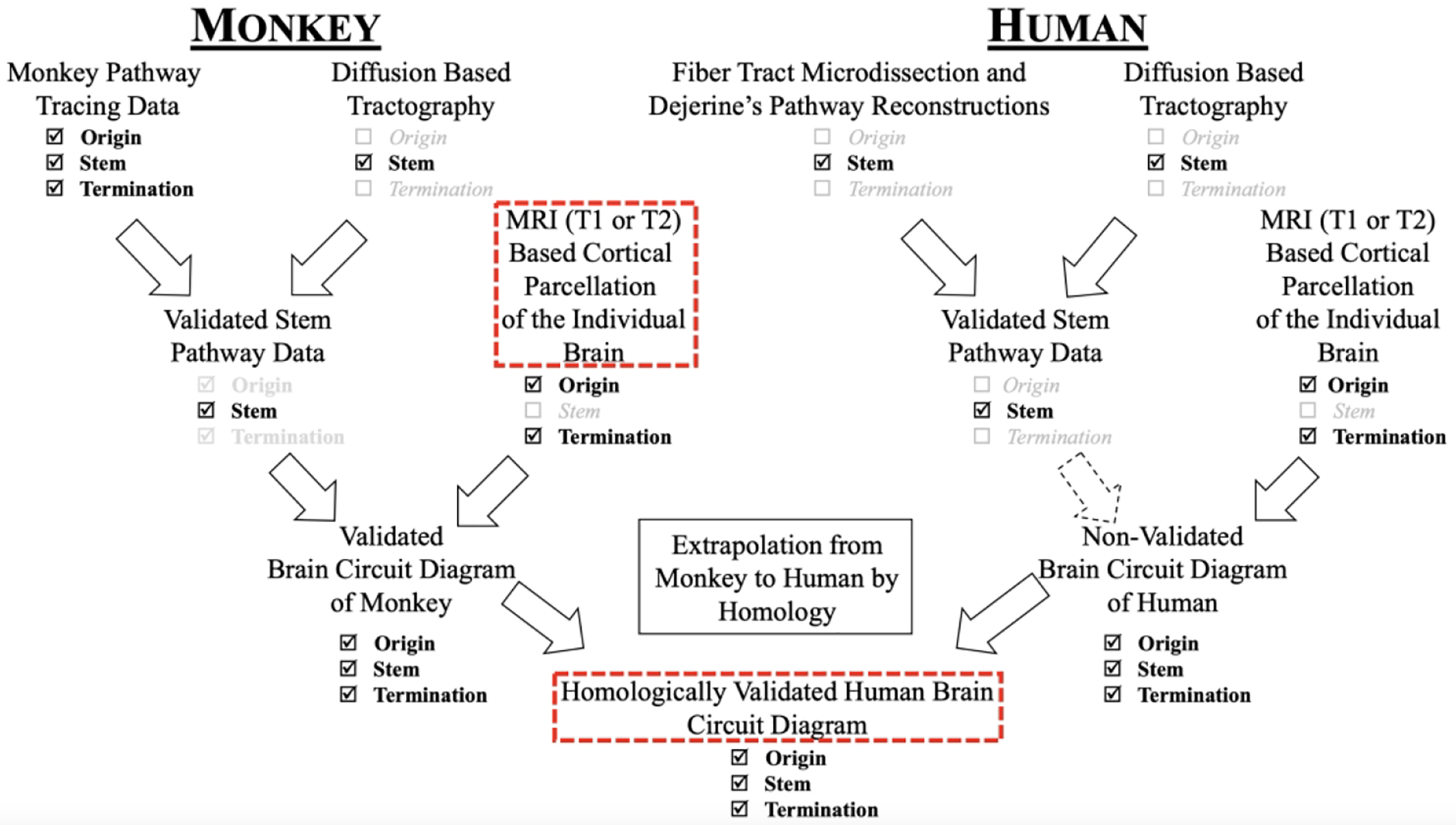
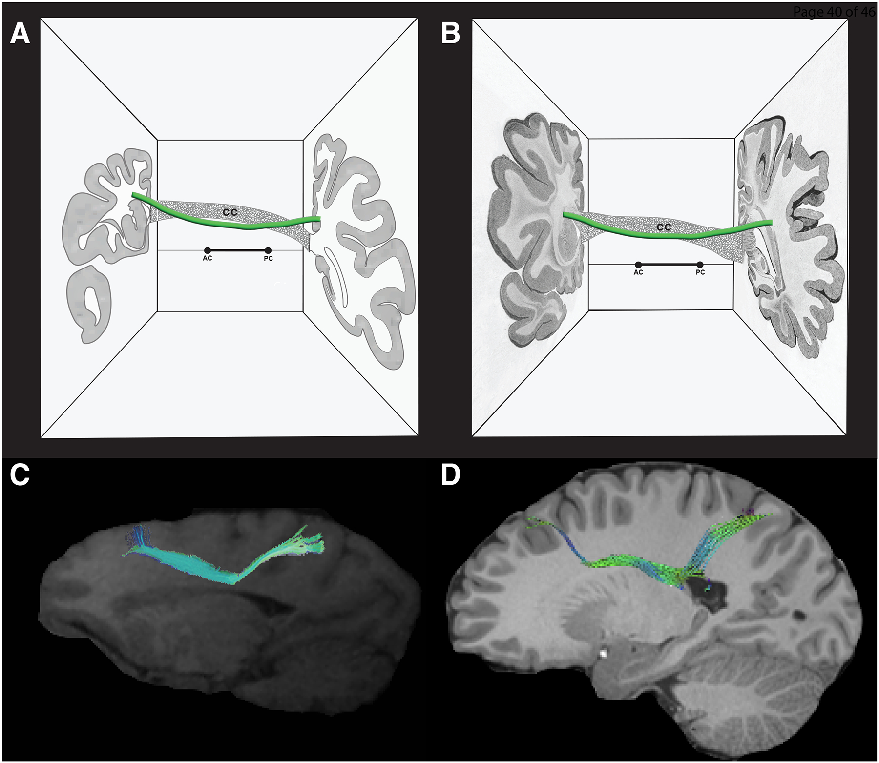
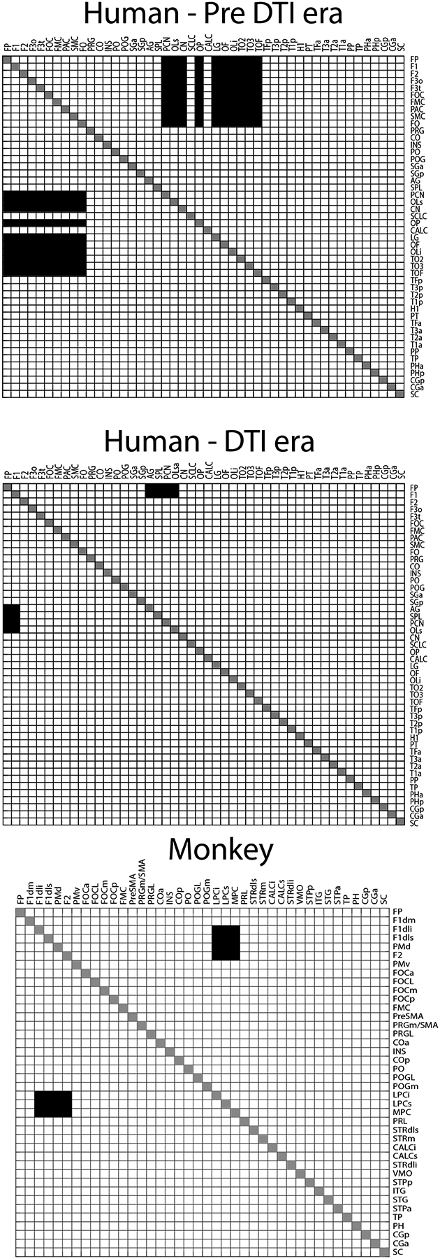
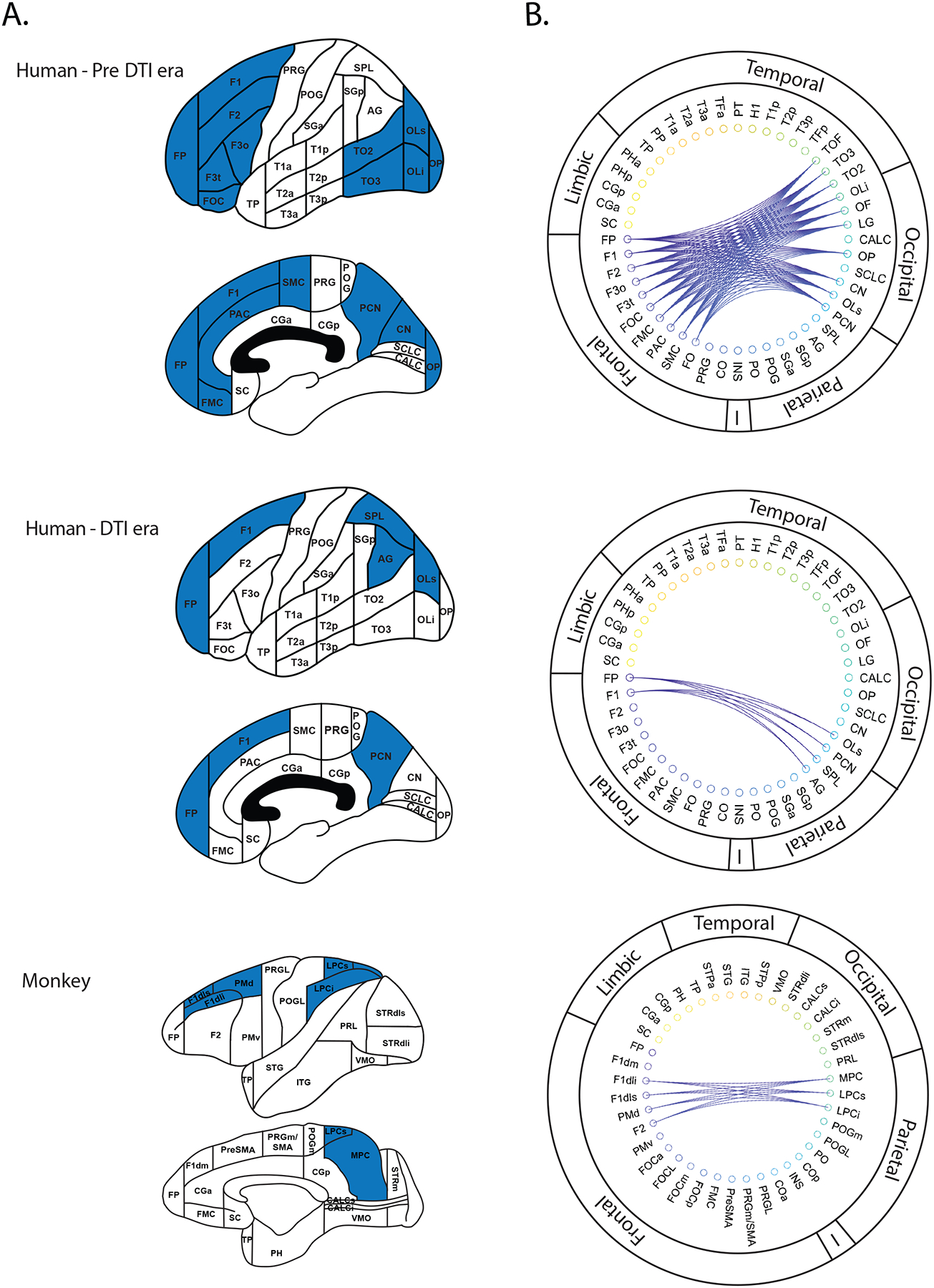
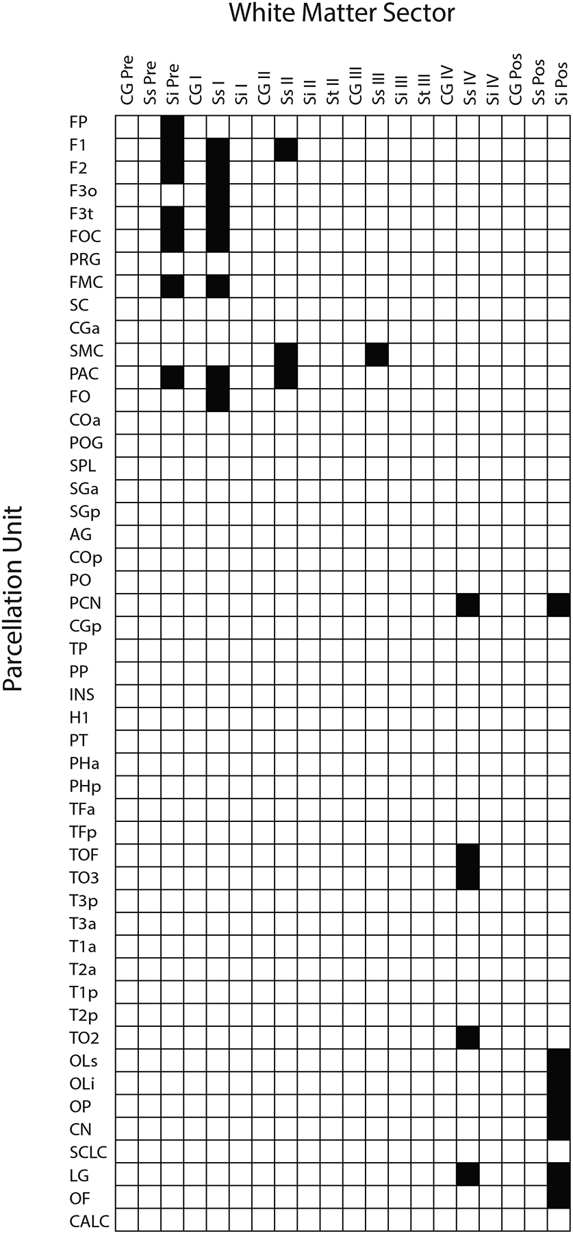
A key set of connections necessary for the most complex brain functions are the long association cortico-cortical fiber tracts. These pathways have been described by the Dejerines and others using post mortem histological or brain dissection techniques. Given methodological limitations, these fiber connections have not been delineated completely in humans. Although the stem portions of fiber tracts have been identified in humans, their precise origins and terminations remain to be determined. By contrast, the origins and terminations as well as the stems of long cortico-cortical association fiber pathways in monkeys have been detailed in the macaque monkey brain using experimental tract tracing methods. Deepak Pandya made major contributions to the delineation of fiber tracts in the monkey brain. In the early 1990s, he compared his observations in monkeys with the original descriptions in humans by the Dejerines. With the advent of diffusion-weighted imaging, Dr. Pandya extended this line of investigation to the human brain with Dr. Nikos Makris. In this translational analysis of long association cortico-cortical fiber tracts, they applied a principle of extrapolation from monkey to human. In the present study, we addressed the reasoning and the complex methodology in translating brain structural connectivity from monkey to human in one cortico-cortical fiber tract, namely the superior fronto-occipital fascicle, which was delineated in both species by Dr. Pandya and colleagues. Furthermore, we represented this information in the form of connectional matrices in the context of the HOA2.0-ComPaRe framework, a homological monkey-to-human translational system used in neuroimaging.
对于最复杂的大脑功能来说,一组关键的连接是长的联合皮质-皮质纤维束。这些通路是由 Dejerines 和其他人使用死后组织学或大脑解剖技术来描述的。由于方法学的限制,这些纤维连接在人类中尚未完全描绘出来。虽然纤维束的干部分已经在人类中被识别出来,但它们的确切起源和终止仍有待确定。相比之下,长皮质-皮质联合纤维通路的起源和终止以及干部分在猴子中已经使用实验追踪方法在猕猴大脑中详细描述。Deepak Pandya 对猴子大脑中的纤维束描绘做出了重大贡献。在 20 世纪 90 年代初,他将自己在猴子身上的观察结果与 Dejerines 最初在人类身上的描述进行了比较。随着扩散加权成像的出现,Pandya 博士与 Nikos Makris 博士一起将这一研究方向扩展到人类大脑。在这项对长联合皮质-皮质纤维束的转化分析中,他们应用了从猴子到人类的外推原理。在本研究中,我们解决了从猴子到人类的大脑结构连接的推理和复杂方法学问题,该纤维束是一个额-枕上束,由 Pandya 博士及其同事在两个物种中描绘。此外,我们在 HOA2.0-ComPaRe 框架的背景下,以连接矩阵的形式表示了这方面的信息,HOA2.0-ComPaRe 是一个用于神经影像学的猴子到人类的同源转化系统。