VIB-KU Leuven Center for Microbiology, Flanders, Belgium.
Laboratory of Molecular Cell Biology, Institute of Botany and Microbiology, KU Leuven, Leuven, Belgium.
mSphere. 2020 Feb 5;5(1):e00981-19. doi: 10.1128/mSphere.00981-19.
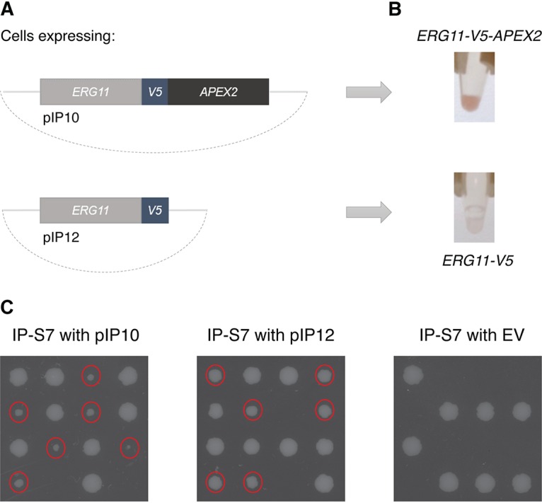
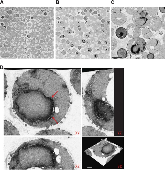
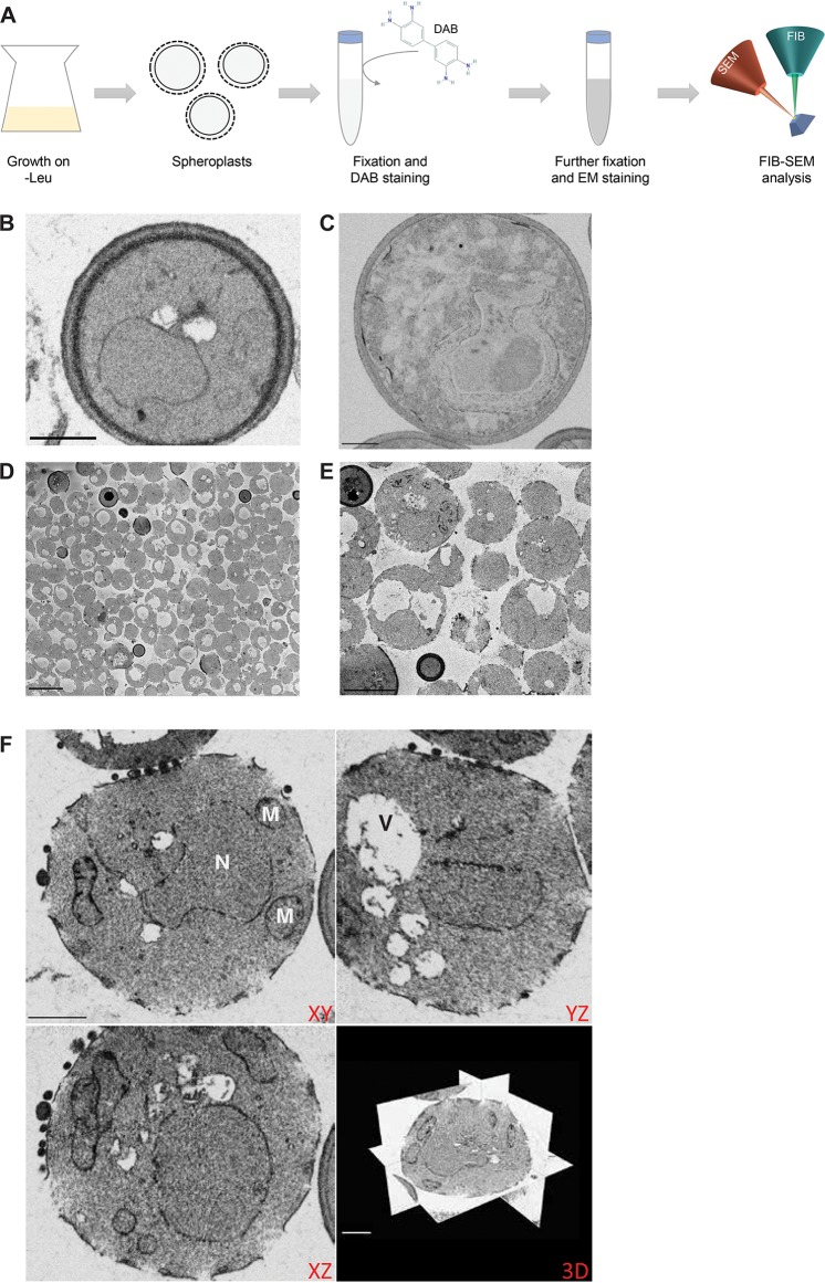
The determination of the exact location of a protein in the cell is essential to the understanding of biological processes. Here, we report for the first time the visualization of a protein of interest in using focused ion beam scanning electron microscopy (FIB-SEM). As a proof of concept, the integral endoplasmic reticulum (ER) membrane protein Erg11 has been C-terminally tagged with APEX2, which is an engineered peroxidase that catalyzes an electron-dense deposition of 3,3'-diaminobenzidine (DAB), as such marking the location of the fused protein of interest in electron microscopic images. As DAB is unable to cross the yeast cell wall to react with APEX2, cell walls have been partly removed by the formation of spheroplasts. This has resulted in a clear electron-dense ER signal for the Erg11 protein using FIB-SEM. With this study, we have validated the use of the APEX2 tag for visualization of yeast proteins in electron microscopy. Furthermore, we have introduced a methodology that enables precise and three-dimensional (3D) localization studies in yeast, with nanometer resolution and without the need for antibody staining. Because of these properties, the described technique can offer valuable information on the molecular functions of studied proteins. With this study, we have validated the use of the APEX2 tag to define the localization of proteins in the model yeast As such, FIB-SEM can identify the exact 3D location of a protein of interest in the cell with nanometer-scale resolution. Such detailed imaging could provide essential information on the elucidation of various biological processes. APEX2, which adds electron density to a fused protein of interest upon addition of the substrate DAB, originally was used in mammalian studies. With this study, we expand its use to protein localization studies in one of the most important models in molecular biology.
确定蛋白质在细胞中的精确位置对于理解生物过程至关重要。在这里,我们首次报道了使用聚焦离子束扫描电子显微镜(FIB-SEM)在酵母中可视化感兴趣的蛋白质。作为概念验证,我们将整合内质网(ER)膜蛋白 Erg11 的 C 端与 APEX2 融合,APEX2 是一种工程过氧化物酶,可催化 3,3'-二氨基联苯胺(DAB)的电子致密沉积,从而在电子显微镜图像中标记融合蛋白的位置。由于 DAB 无法穿过酵母细胞壁与 APEX2 反应,因此通过形成球形体部分去除细胞壁。这导致使用 FIB-SEM 可以清晰地看到 Erg11 蛋白的电子致密 ER 信号。通过这项研究,我们验证了 APEX2 标签在酵母蛋白质电子显微镜可视化中的应用。此外,我们引入了一种方法,该方法能够在酵母中进行精确的三维(3D)定位研究,分辨率达到纳米级,而无需抗体染色。由于这些特性,所描述的技术可以为研究蛋白质的分子功能提供有价值的信息。通过这项研究,我们验证了使用 APEX2 标签来定义模型酵母中蛋白质的定位。因此,FIB-SEM 可以以纳米级分辨率识别细胞中感兴趣的蛋白质的精确 3D 位置。这种详细的成像可以为阐明各种生物学过程提供重要信息。APEX2 在添加底物 DAB 后会向融合蛋白中添加电子密度,最初用于哺乳动物研究。通过这项研究,我们将其用途扩展到分子生物学中最重要的模型之一中的蛋白质定位研究。