Diomede Francesca, Marconi Guya Diletta, Cavalcanti Marcos F X B, Pizzicannella Jacopo, Pierdomenico Sante Donato, Fonticoli Luigia, Piattelli Adriano, Trubiani Oriana
Department of Medical, Oral and Biotechnological Sciences, University "G. D'Annunzio", Chieti-Pescara, 66100 Chieti, Italy.
Biophotonics Laboratory, Nove de Julho University, São Paulo 01506-000, Brazil.
Materials (Basel). 2020 Feb 5;13(3):706. doi: 10.3390/ma13030706.
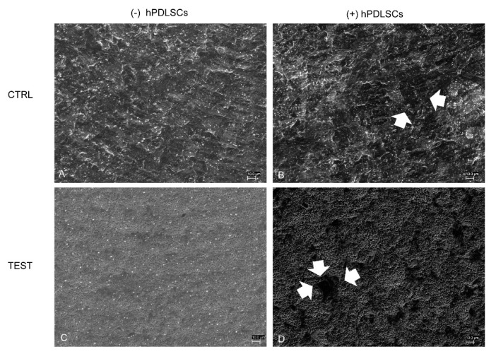
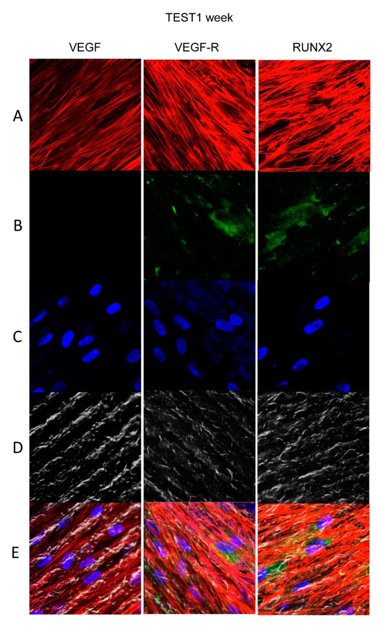
In restorative dentistry, the main implants characteristic is the ability to promote the osseointegration process as the result of interaction between angiogenesis and osteogenesis events. On the other hand, implants cytocompatibility remains a necessary feature for the success of surgery. The purpose of the current study was to investigate the interaction between human periodontal stem cells and two different types of titanium surfaces, to verify their cytocompatibility and cell adhesion ability, and to detect osteogenic and angiogenic markers, trough cell viability assay (MTT), Confocal Laser Scanning Microscopy (CLSM), scanning electron microscopy (SEM), and gene expression (RT-PCR). The titanium surfaces, machined (CTRL) and dual acid etched (TEST), tested in culture with human periodontal ligament stem cells (hPDLSCs), were previously treated in two different ways, in order to evaluate the effects of CTRL and TEST and define the best implant surface. Furthermore, the average surface roughness (Ra) of both titanium surfaces, CTRL and TEST, has been assessed through atomic force microscopy (AFM). The vascular endothelial growth factor (VEGF) and Runt-related transcription factor 2 (RUNX2) expressions have been analyzed by RT-PCR, WB analysis, and confocal laser scanning microscopy. Data evidenced that the different morphology and topography of the TEST disk increased cell growth, cell adhesion, improved osteogenic and angiogenic events, as well osseointegration process. For this reason, the TEST surface was more biocompatible than the CTRL disk surface.
在修复牙科中,植入物的主要特性是能够促进骨整合过程,这是血管生成和成骨事件相互作用的结果。另一方面,植入物的细胞相容性仍然是手术成功的必要特征。本研究的目的是研究人牙周干细胞与两种不同类型钛表面之间的相互作用,验证它们的细胞相容性和细胞粘附能力,并通过细胞活力测定(MTT)、共聚焦激光扫描显微镜(CLSM)、扫描电子显微镜(SEM)和基因表达(RT-PCR)检测成骨和血管生成标志物。用人类牙周膜干细胞(hPDLSCs)在培养中测试的机械加工(CTRL)和双酸蚀刻(TEST)钛表面,之前经过两种不同方式处理,以评估CTRL和TEST的效果并确定最佳植入物表面。此外,通过原子力显微镜(AFM)评估了CTRL和TEST两种钛表面的平均表面粗糙度(Ra)。通过RT-PCR、WB分析和共聚焦激光扫描显微镜分析了血管内皮生长因子(VEGF)和Runt相关转录因子2(RUNX2)的表达。数据表明,TEST盘不同的形态和形貌增加了细胞生长、细胞粘附,改善了成骨和血管生成事件以及骨整合过程。因此,TEST表面比CTRL盘表面具有更高的生物相容性。