ELTE NAP Neuroimmunology Research Group, Department of Biochemistry, Institute of Biology, ELTE Eötvös Loránd University, Budapest, Hungary.
Laboratory of Proteomics, Institute of Biology, ELTE Eötvös Loránd University, Budapest, Hungary.
Cell Mol Life Sci. 2020 Dec;77(24):5243-5258. doi: 10.1007/s00018-020-03468-0. Epub 2020 Feb 7.
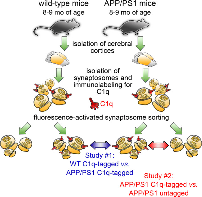
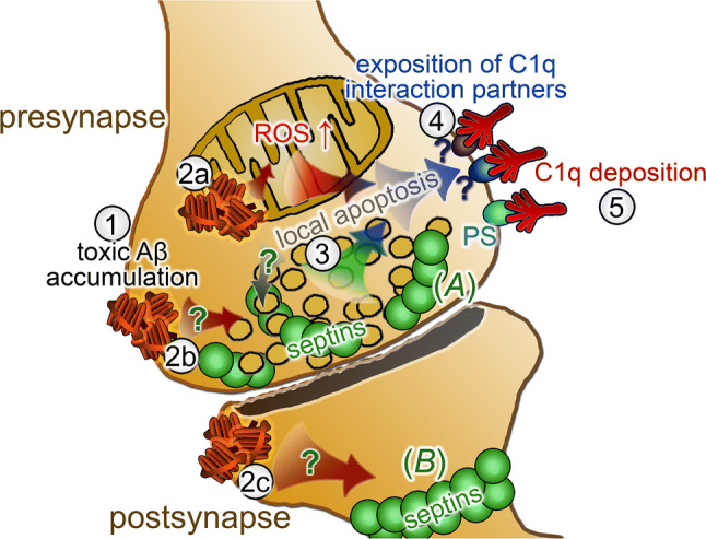
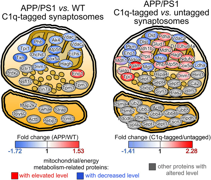
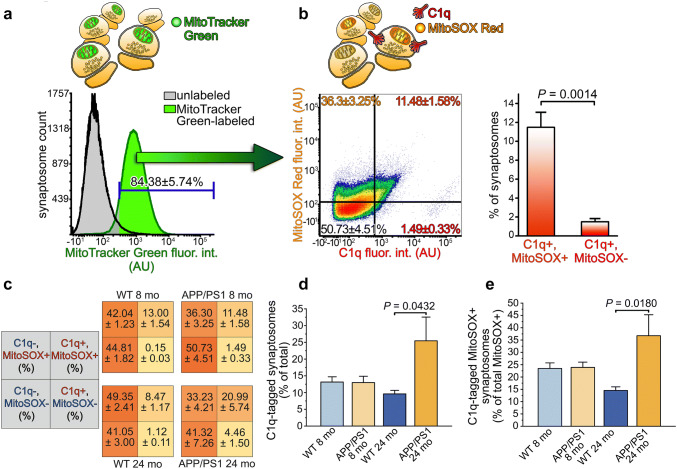
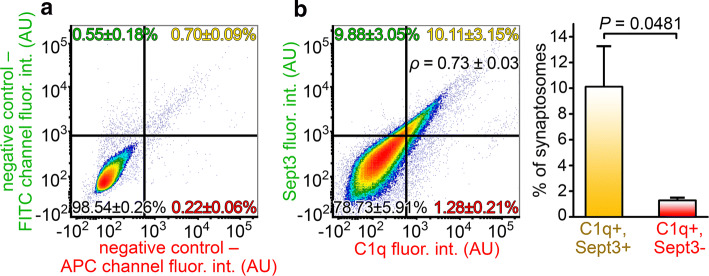
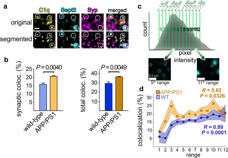
Synaptic functional disturbances with concomitant synapse loss represent central pathological hallmarks of Alzheimer's disease. Excessive accumulation of cytotoxic amyloid oligomers is widely recognized as a key event that underlies neurodegeneration. Certain complement components are crucial instruments of widespread synapse loss because they can tag synapses with functional impairments leading to their engulfment by microglia. However, an exact understanding of the affected synaptic functions that predispose to complement-mediated synapse elimination is lacking. Therefore, we conducted systematic proteomic examinations on synaptosomes prepared from an amyloidogenic mouse model of Alzheimer's disease (APP/PS1). Synaptic fractions were separated according to the presence of the C1q-tag using fluorescence-activated synaptosome sorting and subjected to proteomic comparisons. The results raised the decline of mitochondrial functions in the C1q-tagged synapses of APP/PS1 mice based on enrichment analyses, which was verified using flow cytometry. Additionally, proteomics results revealed extensive alterations in the level of septin protein family members, which are known to dynamically form highly organized pre- and postsynaptic supramolecular structures, thereby affecting synaptic transmission. High-resolution microscopy investigations demonstrated that synapses with considerable amounts of septin-3 and septin-5 show increased accumulation of C1q in APP/PS1 mice compared to the wild-type ones. Moreover, a strong positive correlation was apparent between synaptic septin-3 levels and C1q deposition as revealed via flow cytometry and confocal microscopy examinations. In sum, our results imply that deterioration of synaptic mitochondrial functions and alterations in the organization of synaptic septins are associated with complement-dependent synapse loss in Alzheimer's disease.
突触功能障碍伴突触丢失是阿尔茨海默病的主要病理学特征。细胞毒性淀粉样寡聚物的过度积累被广泛认为是导致神经退行性变的关键事件。某些补体成分是广泛的突触丢失的关键工具,因为它们可以标记功能受损的突触,导致它们被小胶质细胞吞噬。然而,对于导致补体介导的突触消除的受影响的突触功能缺乏确切的理解。因此,我们对来自阿尔茨海默病淀粉样蛋白模型(APP/PS1)的突触体进行了系统的蛋白质组学研究。根据存在 C1q 标记,使用荧光激活的突触体分选将突触部分分离,并进行蛋白质组比较。结果表明,基于富集分析,APP/PS1 小鼠的 C1q 标记突触中的线粒体功能下降,这通过流式细胞术得到了验证。此外,蛋白质组学结果揭示了 septin 蛋白家族成员水平的广泛改变,这些成员已知能够动态形成高度组织化的前突触和后突触超分子结构,从而影响突触传递。高分辨率显微镜研究表明,与野生型相比,APP/PS1 小鼠中具有大量 septin-3 和 septin-5 的突触显示 C1q 的积累增加。此外,通过流式细胞术和共聚焦显微镜检查发现,突触 septin-3 水平与 C1q 沉积之间存在强烈的正相关。总之,我们的结果表明,突触线粒体功能的恶化和突触 septin 的组织改变与阿尔茨海默病中补体依赖性突触丢失有关。