Tamborino Giulia, De Saint-Hubert Marijke, Struelens Lara, Seoane Dayana C, Ruigrok Eline A M, Aerts An, van Cappellen Wiggert A, de Jong Marion, Konijnenberg Mark W, Nonnekens Julie
Research in Dosimetric Application, Belgian Nuclear Research Centre (SCK•CEN), Mol, Belgium.
Department of Radiology & Nuclear Medicine, Erasmus MC, Rotterdam, The Netherlands.
EJNMMI Phys. 2020 Feb 10;7(1):8. doi: 10.1186/s40658-020-0276-5.
Survival and linear-quadratic model fitting parameters implemented in treatment planning for targeted radionuclide therapy depend on accurate cellular dosimetry. Therefore, we have built a refined cellular dosimetry model for [Lu]Lu-DOTA-[Tyr]octreotate (Lu-DOTATATE) in vitro experiments, accounting for specific cell morphologies and sub-cellular radioactivity distributions.
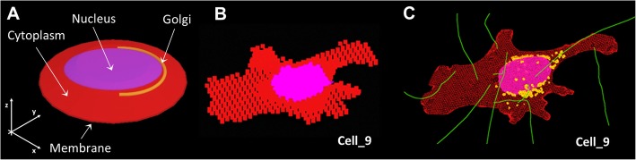
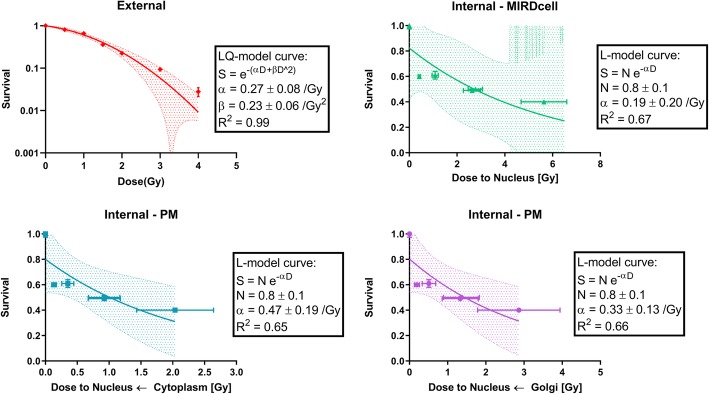
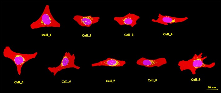
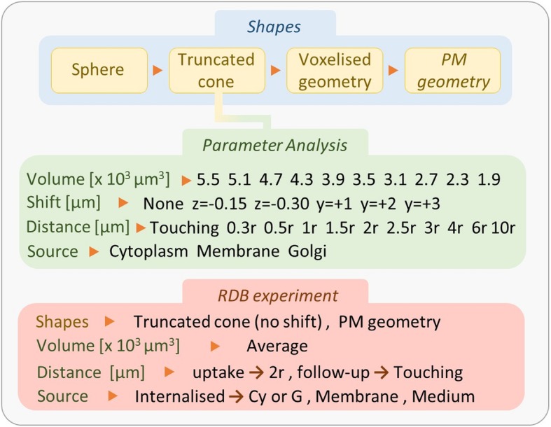
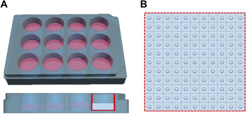
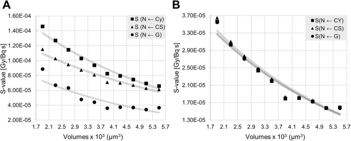
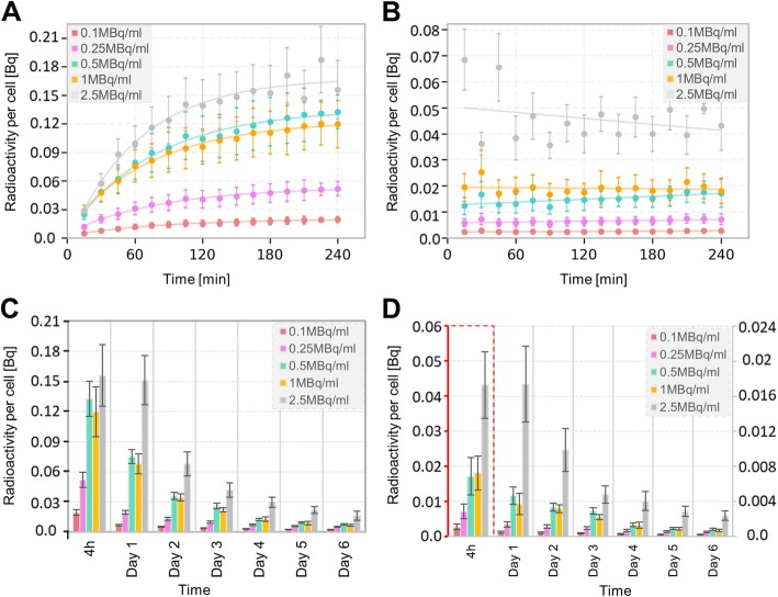
Time activity curves were measured and modeled for medium, membrane-bound, and internalized activity fractions over 6 days. Clonogenic survival assays were performed at various added activities (0.1-2.5 MBq/ml). 3D microscopy images (stained for cytoplasm, nucleus, and Golgi) were used as reference for developing polygonal meshes (PM) in 3DsMax to accurately render the cellular and organelle geometry. Absorbed doses to the nucleus per decay (S values) were calculated for 3 cellular morphologies: spheres (MIRDcell), truncated cone-shaped constructive solid geometry (CSG within MCNP6.1), and realistic PM models, using Geant4-10.03. The geometrical set-up of the clonogenic survival assays was modeled, including dynamic changes in proliferation, proximity variations, and cell death. The absorbed dose to the nucleus by the radioactive source cell (self-dose) and surrounding source cells (cross-dose) was calculated applying the MIRD formalism. Finally, the correlation between absorbed dose and survival fraction was fitted using a linear dose-response curve (high α/β or fast sub-lethal damage repair half-life) for different assumptions, related to cellular shape and localization of the internalized fraction of activity.
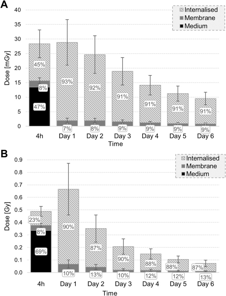
The cross-dose, depending on cell proximity and colony formation, is a minor (15%) contributor to the total absorbed dose. Cellular volume (inverse exponential trend), shape modeling (up to 65%), and internalized source localization (up to + 149% comparing cytoplasm to Golgi) significantly influence the self-dose to nucleus. The absorbed dose delivered to the nucleus during a clonogenic survival assay is 3-fold higher with MIRDcell compared to the polygonal mesh structures. Our cellular dosimetry model indicates that Lu-DOTATATE treatment might be more effective than suggested by average spherical cell dosimetry, predicting a lower absorbed dose for the same cellular survival. Dose-rate effects and heterogeneous dose delivery might account for differences in dose-response compared to x-ray irradiation.
Our results demonstrate that modeling of cellular and organelle geometry is crucial to perform accurate in vitro dosimetry.
在靶向放射性核素治疗的治疗计划中实施的生存和线性二次模型拟合参数取决于精确的细胞剂量测定。因此,我们构建了一个用于[Lu]Lu-DOTA-[Tyr]奥曲肽(Lu-DOTATATE)体外实验的精细细胞剂量测定模型,该模型考虑了特定的细胞形态和亚细胞放射性分布。
测量并模拟了6天内培养基、膜结合和内化活性部分的时间-活性曲线。在不同添加活性(0.1 - 2.5 MBq/ml)下进行克隆形成存活测定。3D显微镜图像(用细胞质、细胞核和高尔基体染色)用作在3DsMax中开发多边形网格(PM)的参考,以准确呈现细胞和细胞器的几何形状。使用Geant4-10.03为3种细胞形态计算每个衰变对细胞核的吸收剂量(S值):球体(MIRDcell)、截头圆锥形建设性实体几何形状(MCNP6.1中的CSG)和真实的PM模型。对克隆形成存活测定的几何设置进行建模,包括增殖的动态变化、邻近度变化和细胞死亡。应用MIRD形式计算放射性源细胞对细胞核的吸收剂量(自剂量)和周围源细胞的吸收剂量(交叉剂量)。最后,针对与细胞形状和内化活性部分的定位相关的不同假设,使用线性剂量响应曲线(高α/β或快速亚致死损伤修复半衰期)拟合吸收剂量与存活分数之间的相关性。
取决于细胞邻近度和集落形成的交叉剂量对总吸收剂量的贡献较小(15%)。细胞体积(反指数趋势)、形状建模(高达65%)和内化源定位(将细胞质与高尔基体比较时高达 + 149%)对细胞核的自剂量有显著影响。与多边形网格结构相比,在克隆形成存活测定期间传递到细胞核的吸收剂量,MIRDcell高出3倍。我们的细胞剂量测定模型表明,Lu-DOTATATE治疗可能比平均球形细胞剂量测定所表明的更有效,但对于相同的细胞存活预测吸收剂量更低。与X射线照射相比,剂量率效应和异质剂量传递可能是剂量响应差异的原因。
我们的结果表明,细胞和细胞器几何形状的建模对于进行准确的体外剂量测定至关重要。