School of Medical Sciences.
Physics Institute.
Nucl Med Commun. 2020 Apr;41(4):377-382. doi: 10.1097/MNM.0000000000001165.
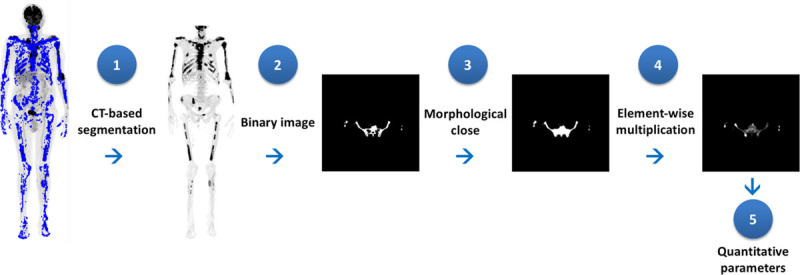
Quantifications in nuclear medicine are occasionally limited by the lack of standardization for defining volumes of interest (VOIs) on functional images. In the present article, we propose the use of computed tomography (CT)-based skeletal segmentation to determine anatomically the VOI in order to calculate quantitative parameters of fluorine 18 fluorodeoxyglucose (F-FDG) PET/CT images from patients with multiple myeloma.
We evaluated 101 whole-body F-FDG PET/CTs of 58 patients with multiple myeloma. An initial subjective visual analysis of the PET images was used to classify the bone involvement as negative/mild, moderate, or marked. Then, a fully automated CT-based segmentation of the skeleton was performed on PET images. The maximum, mean, and SD of the standardized uptake values (SUVmax, SUVmean, and SDSUV) were calculated for bone tissue and compared with the visual analysis.
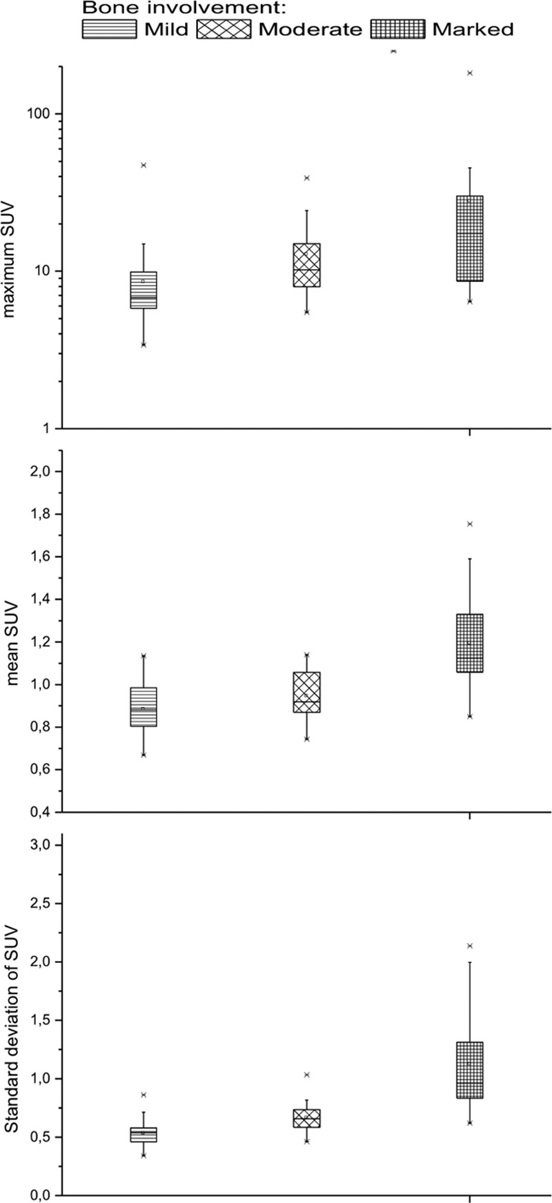
Forty-five (44.5%), 32 (31.7%), and 24 (23.8%) PET images were, respectively, classified as negative/mild, moderate, or marked bone involvement. All quantitative parameters were significantly related to the visual assessment of bone involvement. This association was stronger for the SUVmean [odds ratio (OR): 10.52 (95% confidence interval (CI), 5.68-19.48); P < 0.0001] and for the SDSUV [OR: 5.58 (95% CI, 3.31-9.42); P < 0.001) than for the SUVmax [OR: 1.01 (95% CI, 1.003-1.022); P = 0.003].
CT-based skeletal segmentation allows for automated and therefore reproducible calculation of PET quantitative parameters of bone involvement in patients with multiple myeloma. Using this method, the SUVmean and its respective SD correlated better with the visual analysis of F-FDG PET images than SUVmax. Its value in staging and evaluating therapy response needs to be evaluated.
在核医学中,由于缺乏定义功能图像中感兴趣区(VOI)体积的标准化方法,定量分析有时会受到限制。在本文中,我们提出使用基于计算机断层扫描(CT)的骨骼分割来从解剖学上确定 VOI,以便计算多发性骨髓瘤患者氟 18 氟脱氧葡萄糖(F-FDG)PET/CT 图像的定量参数。
我们评估了 58 例多发性骨髓瘤患者的 101 例全身 F-FDG PET/CT。首先对 PET 图像进行主观视觉分析,将骨受累分为阴性/轻度、中度或重度。然后,对 PET 图像进行全自动基于 CT 的骨骼分割。计算骨组织的标准化摄取值最大值(SUVmax)、平均值(SUVmean)和标准差(SDSUV),并与视觉分析进行比较。
45 例(44.5%)、32 例(31.7%)和 24 例(23.8%)PET 图像分别被归类为阴性/轻度、中度或重度骨受累。所有定量参数与骨受累的视觉评估均显著相关。SUVmean 与骨受累的视觉评估相关性更强[优势比(OR):10.52(95%置信区间(CI),5.68-19.48);P<0.0001],SDSUV 与骨受累的视觉评估相关性也更强[OR:5.58(95% CI,3.31-9.42);P<0.001],而 SUVmax 与骨受累的视觉评估相关性较弱[OR:1.01(95% CI,1.003-1.022);P=0.003]。
基于 CT 的骨骼分割允许自动且可重复地计算多发性骨髓瘤患者骨骼受累的 PET 定量参数。使用该方法,SUVmean 及其相应的 SD 与 F-FDG PET 图像的视觉分析相关性优于 SUVmax。其在分期和评估治疗反应中的价值需要进一步评估。