Department of Neuroanatomy and Molecular Brain Research, Institute of Anatomy, Ruhr-University of Bochum, Bochum, Germany.
Section for Computational Sensomotorics, Department of Cognitive Neurology, Hertie-Institute for Clinical Brain Research and Centre for Integrative Neuroscience, University of Tübingen, Tübingen, Germany.
Eur Arch Psychiatry Clin Neurosci. 2020 Oct;270(7):819-828. doi: 10.1007/s00406-020-01107-0. Epub 2020 Feb 15.
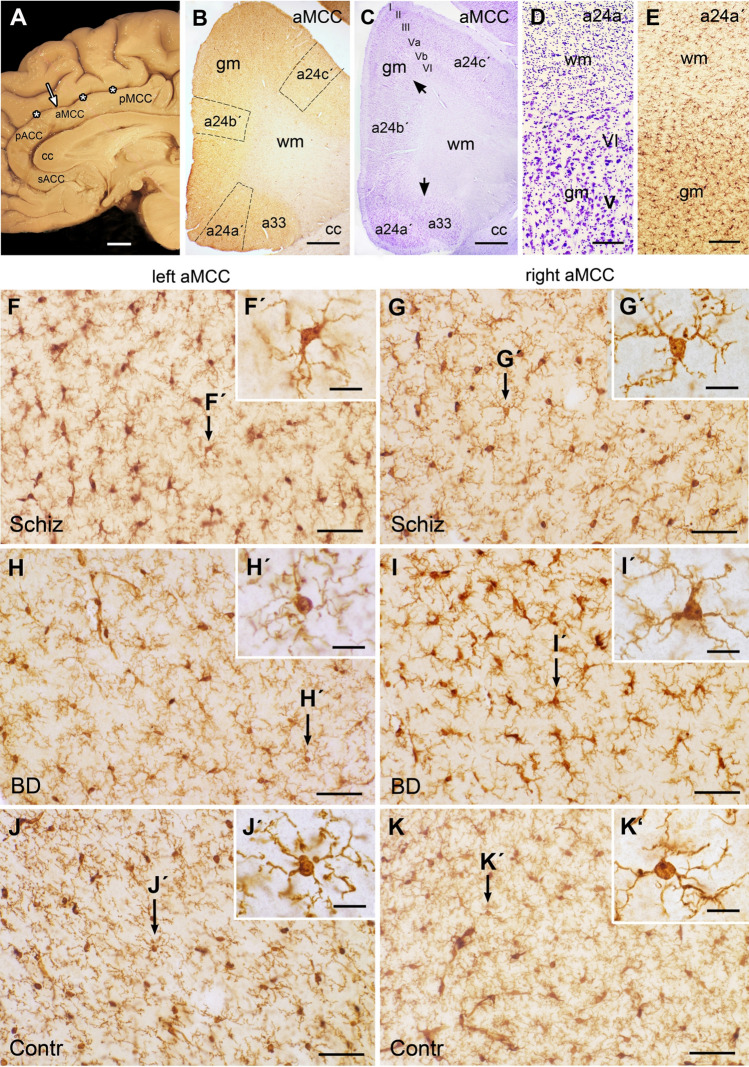
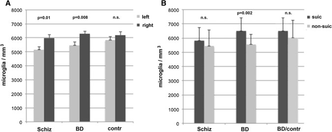
There is increasing evidence from genetic, biochemical, pharmacological, neuroimaging and post-mortem studies that immunological dysregulation plays a crucial role in the pathogenesis of psychoses. The involvement of microglia in schizophrenia and bipolar disorder (BD) has remained controversial, however, since results from various post-mortem studies are still inconclusive. Here, we analyzed the estimated density of microglia of age-matched individuals with schizophrenia (n = 17), BD (n = 13), and non-psychiatric control subjects (n = 17) in the anterior midcingulate cortex (aMCC), a brain area putatively involved in the pathogenesis of psychoses, using ionized calcium binding adaptor molecule 1 (Iba1)-immunohistochemistry. The microglial cells displayed a homogenously distributed Iba1-staining pattern in the aMCC with slightly varying activation states in all three groups. The estimated microglial densities did not differ significantly between individuals with schizophrenia, BD and control subjects. Remarkably, when both hemispheres were investigated separately within the three groups, the density was significantly lateralized towards the right aMCC in schizophrenia (p = 0.01) and-even more evident-in BD subjects (p = 0.008). This left-right lateralization was not observed in the control group (p = 0.52). Of note, microglial density was significantly lower in BD individuals who did not commit suicide compared with BD individuals who died from suicide (p = 0.002). This difference was not observed between individuals with BD who committed suicide and controls. The results, tentatively interpreted, suggest a hitherto unknown increased lateralization of microglial density to the right hemisphere in both psychiatric groups. If confirmed in independent samples, lateralization should be considered in all post-mortem studies on microglia. Density differences between suicide and non-suicide individuals needs further elucidation.
越来越多的证据表明,免疫失调在精神病的发病机制中起着至关重要的作用,这些证据来自遗传、生化、药理学、神经影像学和尸检研究。然而,小胶质细胞在精神分裂症和双相情感障碍(BD)中的作用仍然存在争议,因为各种尸检研究的结果仍然没有定论。在这里,我们使用离子钙结合衔接分子 1(Iba1)免疫组织化学分析了年龄匹配的精神分裂症(n=17)、BD(n=13)和非精神病对照受试者(n=17)在前扣带皮层(aMCC)中的小胶质细胞估计密度,该脑区被认为与精神病的发病机制有关。在所有三组中,小胶质细胞均显示出均匀分布的 Iba1 染色模式,其激活状态略有不同。精神分裂症、BD 和对照组个体之间的小胶质细胞密度没有显著差异。值得注意的是,当在三组内分别研究两个半球时,精神分裂症患者的右侧 aMCC 密度显著偏向右侧(p=0.01),在 BD 患者中更为明显(p=0.008)。对照组中未观察到这种左右偏侧化(p=0.52)。值得注意的是,与自杀的 BD 患者相比,未自杀的 BD 患者的小胶质细胞密度显著降低(p=0.002)。BD 患者中自杀和对照组之间没有观察到这种差异。这些结果提示,在两个精神科群体中,小胶质细胞密度的右偏侧化程度增加,这是迄今为止未知的。如果在独立样本中得到证实,那么在所有关于小胶质细胞的尸检研究中都应考虑偏侧化。自杀和非自杀个体之间的密度差异需要进一步阐明。