Weissenberger M, Weissenberger M H, Gilbert F, Groll J, Evans C H, Steinert A F
Department of Orthopaedic Surgery, König-Ludwig-Haus, Center for Musculoskeletal Research, Julius-Maximilians-University, Brettreichstrasse 11, D-97074, Würzburg, Germany.
Department of Pathology, Caritas-Hospital, Bad Mergentheim, Germany.
BMC Musculoskelet Disord. 2020 Feb 17;21(1):109. doi: 10.1186/s12891-020-3137-4.
Mesenchymal stem cell (MSC) based-treatments of cartilage injury are promising but impaired by high levels of hypertrophy after chondrogenic induction with several bone morphogenetic protein superfamily members (BMPs). As an alternative, this study investigates the chondrogenic induction of MSCs via adenoviral gene-delivery of the transcription factor SOX9 alone or in combination with other inducers, and comparatively explores the levels of hypertrophy and end stage differentiation in a pellet culture system in vitro.
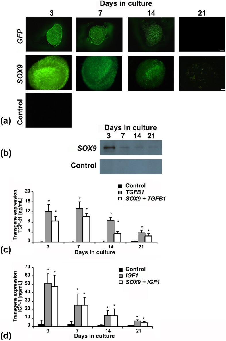
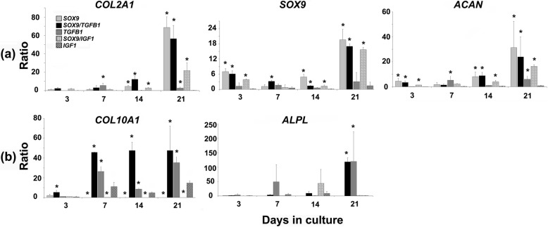
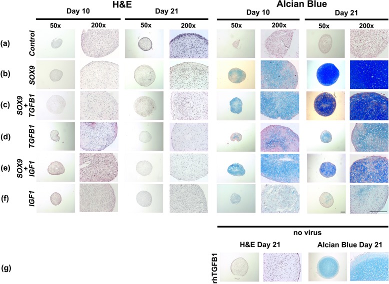
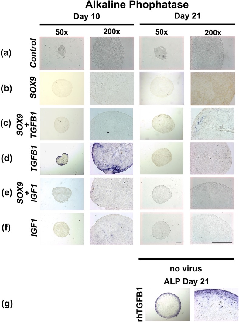
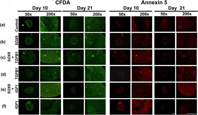
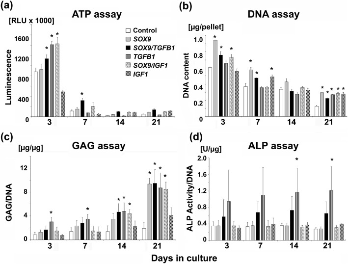
First generation adenoviral vectors encoding SOX9, TGFB1 or IGF1 were used alone or in combination to transduce human bone marrow-derived MSCs at 5 × 10 infectious particles/cell. Thereafter cells were placed in aggregates and maintained for three weeks in chondrogenic medium. Transgene expression was determined at the protein level (ELISA/Western blot), and aggregates were analysed histologically, immunohistochemically, biochemically and by RT-PCR for chondrogenesis and hypertrophy.
SOX9 cDNA was superior to that encoding TGFB1, the typical gold standard, as an inducer of chondrogenesis in primary MSCs as evidenced by improved lacuna formation, proteoglycan and collagen type II staining, increased levels of GAG synthesis, and expression of mRNAs associated with chondrogenesis. Moreover, SOX9 modified aggregates showed a markedly lower tendency to progress towards hypertrophy, as judged by expression of the hypertrophy markers alkaline phosphatase, and collagen type X at the mRNA and protein levels.
Adenoviral SOX9 gene transfer induces chondrogenic differentiation of human primary MSCs in pellet culture more effectively than TGFB1 gene transfer with lower levels of chondrocyte hypertrophy after 3 weeks of in vitro culture. Such technology might enable the formation of more stable hyaline cartilage repair tissues in vivo.
基于间充质干细胞(MSC)的软骨损伤治疗前景广阔,但在用几种骨形态发生蛋白超家族成员(BMP)进行软骨形成诱导后,会因高水平的肥大而受到影响。作为一种替代方法,本研究通过单独或与其他诱导剂联合腺病毒基因递送转录因子SOX9来研究MSC的软骨形成诱导,并在体外微团培养系统中比较探索肥大和终末分化水平。
使用编码SOX9、TGFB1或IGF1的第一代腺病毒载体单独或联合以5×10感染颗粒/细胞转导人骨髓来源的MSC。此后,将细胞聚集成团,并在软骨形成培养基中维持三周。在蛋白质水平(ELISA/蛋白质免疫印迹)测定转基因表达,并对微团进行组织学、免疫组织化学、生物化学分析以及通过RT-PCR分析软骨形成和肥大情况。
作为原代MSC软骨形成的诱导剂,SOX9 cDNA优于编码TGFB1的cDNA(典型的金标准),这通过改善陷窝形成、蛋白聚糖和II型胶原染色、增加糖胺聚糖合成水平以及与软骨形成相关的mRNA表达得以证明。此外,通过在mRNA和蛋白质水平上肥大标志物碱性磷酸酶和X型胶原的表达判断,经SOX9修饰的微团向肥大发展的趋势明显更低。
腺病毒SOX9基因转移比TGFB1基因转移更有效地诱导人原代MSC在微团培养中的软骨形成分化,且在体外培养3周后软骨细胞肥大水平更低。这种技术可能使体内形成更稳定的透明软骨修复组织。