Bhoora Sachin, Pather Yuvelia, Marais Sumari, Punchoo Rivak
Department of Chemical Pathology, Faculty of Health Sciences, University of Pretoria, Pretoria 0083, South Africa.
Department of Physiology, Faculty of Health Sciences, University of Pretoria, Pretoria 0083, South Africa.
Med Sci (Basel). 2020 Feb 13;8(1):12. doi: 10.3390/medsci8010012.
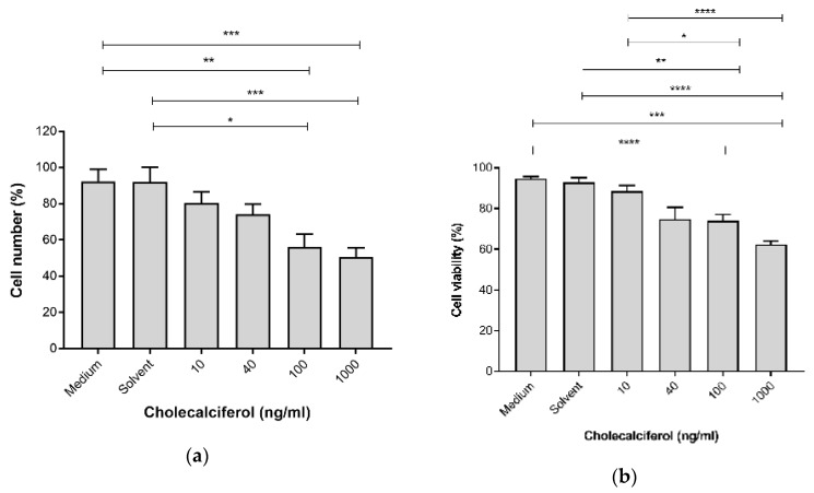
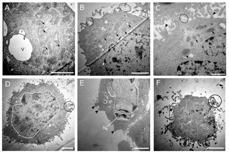
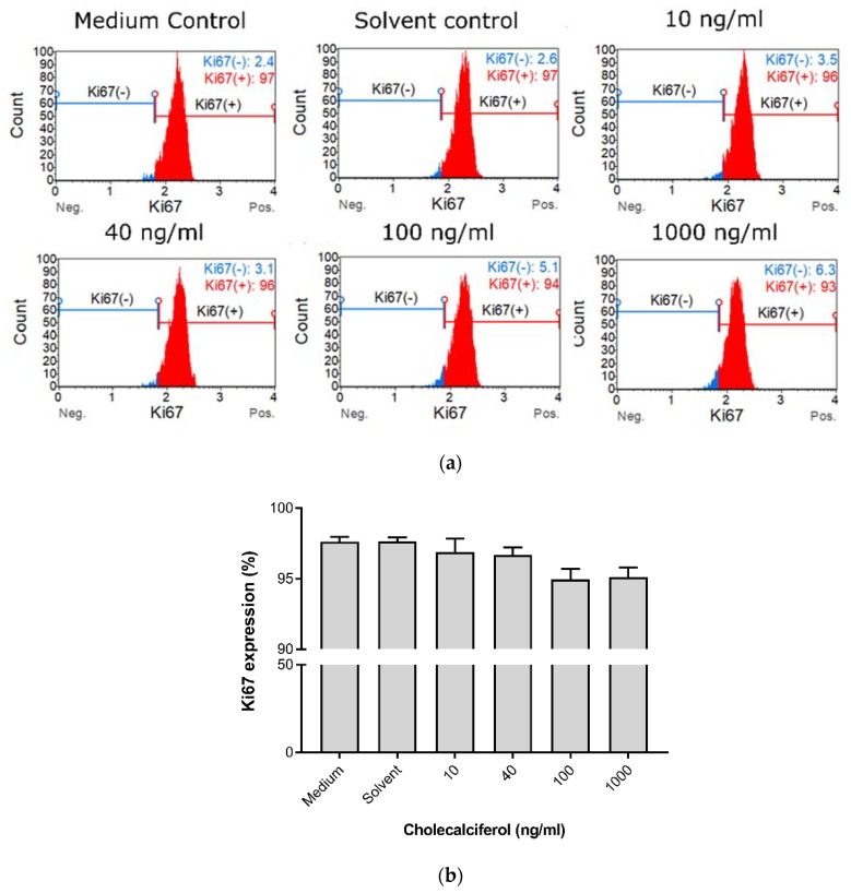
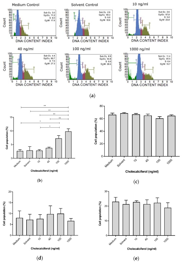
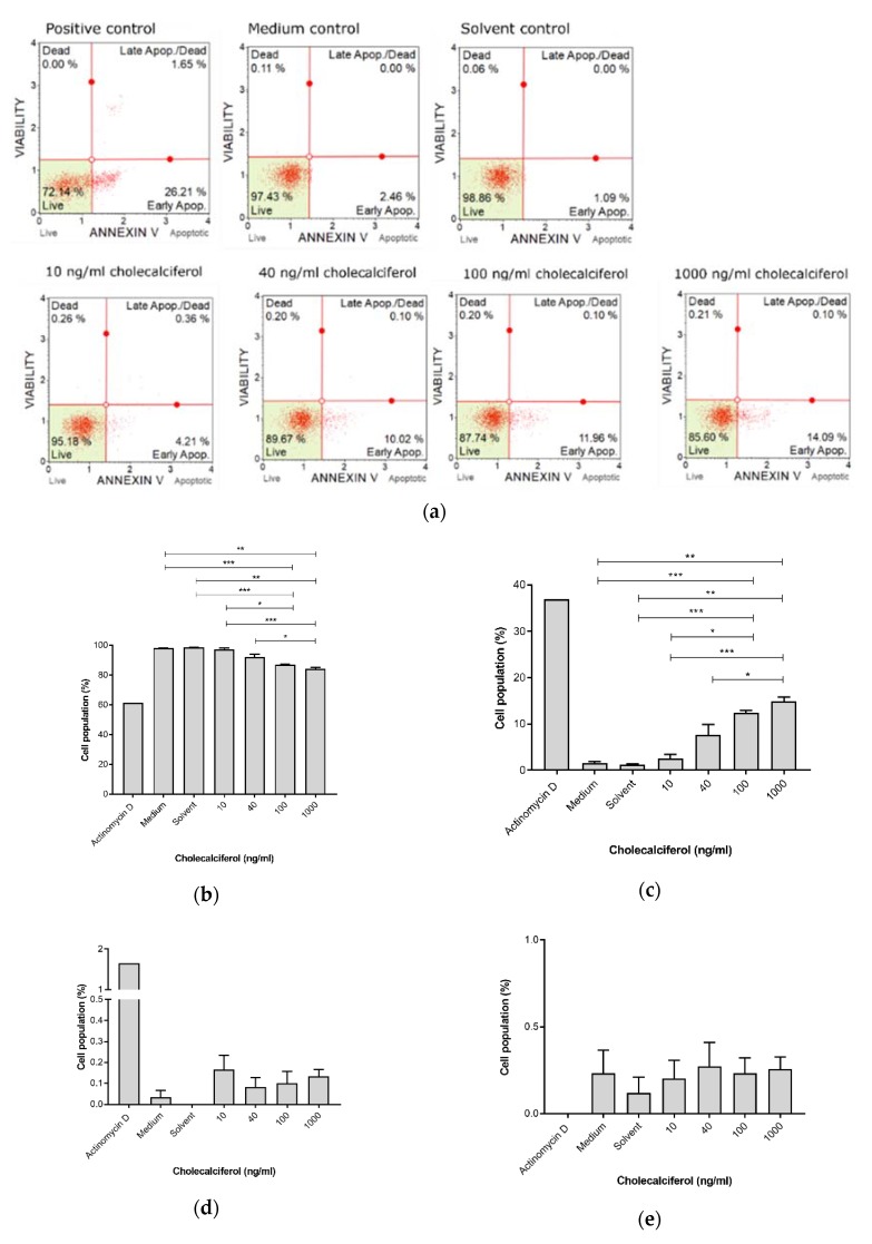
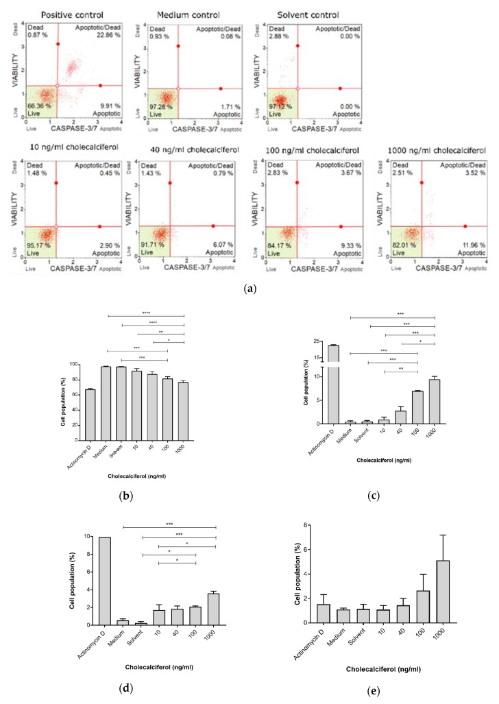
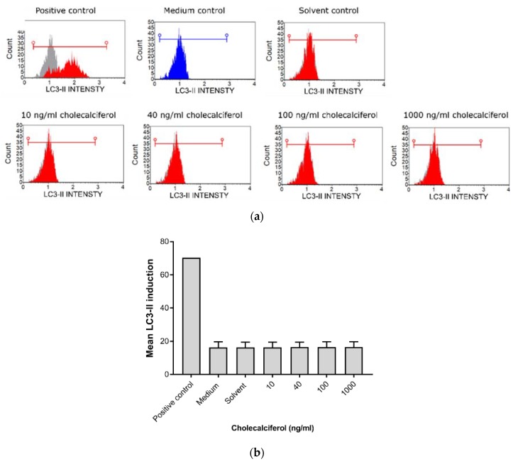
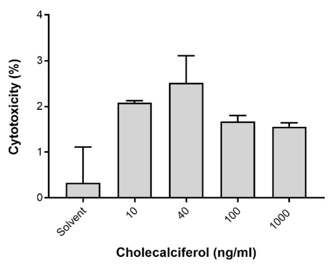
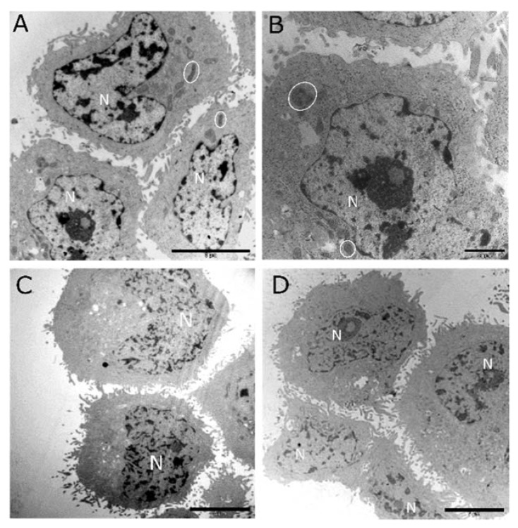
Vitamin D has displayed anti-cancer actions in numerous in vitro studies. Here, we investigated the anti-cancer actions of cholecalciferol, a vitamin D precursor, on a metastatic cervical cancer cell line, namely, CaSki. Experimental cultures were incubated for 72 h and treated with cholecalciferol (10-1000 ng/mL). In the present study, cell count, viability, proliferation and cell cycle were analyzed by a crystal violet assay, trypan blue assay, Ki67 proliferation, and a cell cycle assay, respectively. Biomarkers of apoptosis, necrosis, and autophagic cell death were measured by the Caspase 3/7 and Annexin V/7-AAD Muse™ assays, a LC3-II assay, and a lactate dehydrogenase release assay, respectively. The ultrastructural features of cell death were assessed by transmission electron microscopy. A statistical analysis was performed using a one-way ANOVA and Bonferroni's post-hoc analysis test, and < 0.05 is considered statistically significant here. The results identify statistical decreases in cell count and viability at high-dose treatments (100 and 1000 ng/mL). In addition, significant increases in apoptotic biochemical markers and apoptotic ultrastructure are shown to be present at high-dose treatments. In conclusion, high-dose cholecalciferol treatments inhibit cell count and viability, which are both mediated by apoptotic induction in the CaSki cell line.
维生素D在众多体外研究中已显示出抗癌作用。在此,我们研究了维生素D前体胆钙化醇对转移性宫颈癌细胞系CaSki的抗癌作用。将实验培养物孵育72小时,并用胆钙化醇(10 - 1000 ng/mL)处理。在本研究中,分别通过结晶紫测定、台盼蓝测定、Ki67增殖和细胞周期测定来分析细胞计数、活力、增殖和细胞周期。分别通过Caspase 3/7和膜联蛋白V/7-AAD Muse™测定、LC3-II测定和乳酸脱氢酶释放测定来测量凋亡、坏死和自噬性细胞死亡的生物标志物。通过透射电子显微镜评估细胞死亡的超微结构特征。使用单因素方差分析和Bonferroni事后分析检验进行统计分析,在此处<0.05被认为具有统计学意义。结果表明,高剂量处理(100和1000 ng/mL)时细胞计数和活力有统计学意义的下降。此外,高剂量处理时凋亡生化标志物和凋亡超微结构有显著增加。总之,高剂量胆钙化醇处理可抑制细胞计数和活力,这两者均由CaSki细胞系中的凋亡诱导介导。