Järvinen Jyri, Niinimäki Jaakko, Karppinen Jaro, Takalo Reijo, Haapea Marianne, Tervonen Osmo
Department of Diagnostic Radiology, Oulu University Hospital, Oulu, Finland.
Medical Research Center Oulu, University of Oulu and Oulu University Hospital, Oulu, Finland.
Eur J Radiol Open. 2020 Feb 10;7:100222. doi: 10.1016/j.ejro.2020.100222. eCollection 2020.
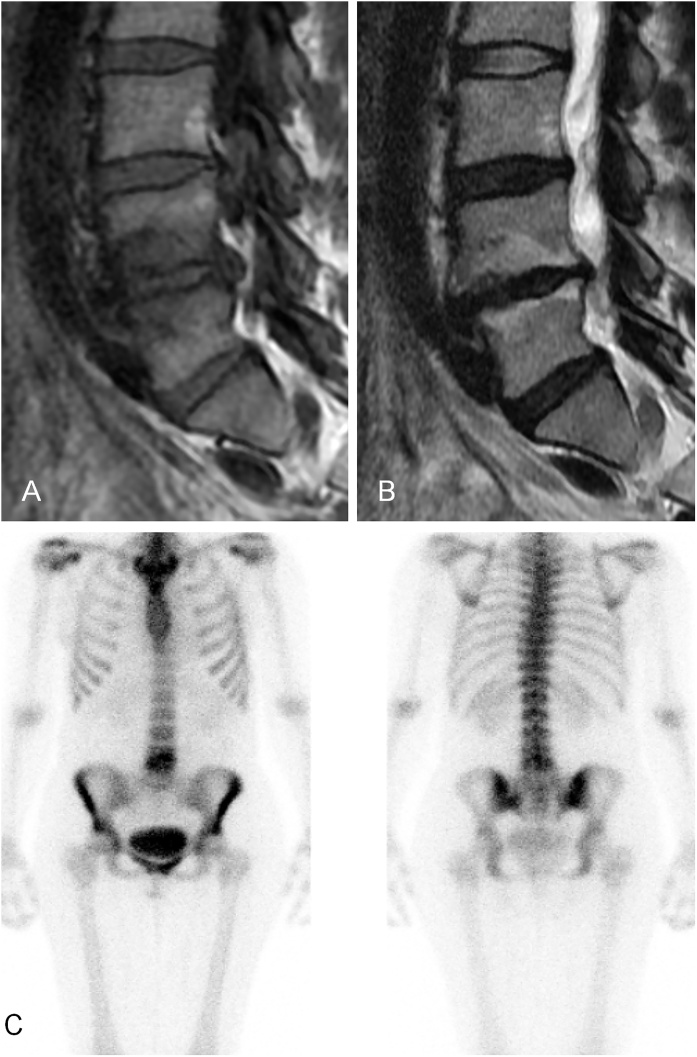
Our purpose was to evaluate whether Modic changes (MC) revealed in lumbar MRI are associated with increased tracer uptake shown in bone scintigraphy. To our knowledge, this has not previously been studied.
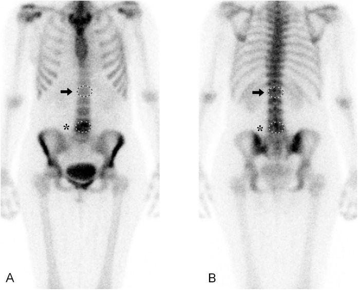
We included patients with MC shown in lumbar MRI and bone scintigraphy performed within six months before or after MRI. Exclusion criteria included metastasis and other specific lesions in the area of interest such as discitis, tumors or fractures. We compared the level and type of MC to the degree of tracer uptake shown in bone scintigraphy. Tracer uptake was assessed both visually and quantitatively. We calculated the lesion-to-normal-bone ratios between the MC area with increased tracer uptake and the vertebra with normal tracer uptake. We used linear mixed models in statistical analyses.
Our study sample consisted of 93 patients (aged 37-86) with 299 MC (28 Type 1 (M1), 50 mixed Type 1/2 (M1/2), 3 mixed Type 1/3 (M1/3), 211 Type 2 (M2), 6 mixed Type 2/3 (M2/3), and 1 Type 3 (M3)). Of all the MC, 26 (93 %) M1, 34 (64 %) in the combined M1/2 and M1/3 group, and 11 (5 %) in the combined M2, M2/3 and M3 group showed increased tracer uptake. The mean lesion-to-normal-bone ratio was higher for lesions with a Type 1 component (M1, M1/2 and M1/3) than for other types, at 1.55 (SD 0.16) for M1; 1.44 (SD 0.21) for combined M1/2 and M1/3; and 1.28 (SD 0.11) for combined M2, M2/3 and M3; p = 0.001).
In most cases, MC with a Type 1 component showed increased tracer uptake in bone scintigraphy. This indicates that bone turnover is accelerated in the M1 area.
我们的目的是评估腰椎磁共振成像(MRI)显示的Modic改变(MC)是否与骨闪烁显像中示踪剂摄取增加相关。据我们所知,此前尚未有过相关研究。
我们纳入了腰椎MRI显示有MC且在MRI前后六个月内进行了骨闪烁显像的患者。排除标准包括转移瘤以及感兴趣区域的其他特定病变,如椎间盘炎、肿瘤或骨折。我们将MC的程度和类型与骨闪烁显像中示踪剂摄取程度进行比较。示踪剂摄取通过视觉和定量方式进行评估。我们计算了示踪剂摄取增加的MC区域与示踪剂摄取正常的椎体之间的病变与正常骨的比值。我们在统计分析中使用线性混合模型。
我们的研究样本包括93例患者(年龄37 - 86岁),共有299处MC(28处1型(M1)、50处1/2混合型(M1/2)、3处1/3混合型(M1/3)、211处2型(M2)、6处2/3混合型(M2/3)和1处3型(M3))。在所有MC中,26处(93%)M1、M1/2和M1/3组合组中的34处(64%)以及M2、M2/3和M3组合组中的11处(5%)显示示踪剂摄取增加。具有1型成分的病变(M1、M1/2和M1/3)的平均病变与正常骨比值高于其他类型,M1为1.55(标准差0.16);M1/2和M1/3组合为1.44(标准差0.21);M2、M2/3和M3组合为1.28(标准差0.11);p = 0.001)。
在大多数情况下,具有1型成分的MC在骨闪烁显像中显示示踪剂摄取增加。这表明M1区域的骨转换加速。