Önder Ömer, Azizova Aynur, Durhan Gamze, Elibol Funda Dinç, Akpınar Meltem Gülsün, Demirkazık Figen
Department of Radiology, School of Medicine, Hacettepe University, 06100, Ankara, Turkey.
Department of Radiology, School of Medicine, Muğla Sıtkı Koçman University, 48000, Muğla, Turkey.
Insights Imaging. 2020 Feb 18;11(1):27. doi: 10.1186/s13244-019-0834-3.
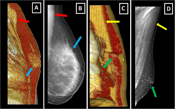
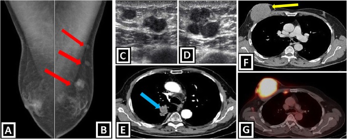
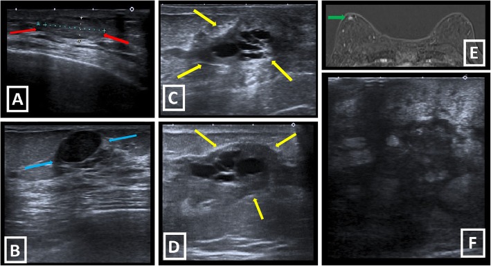
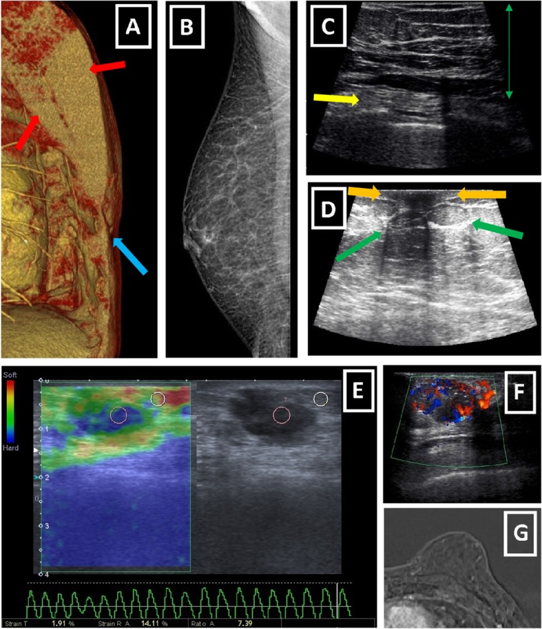
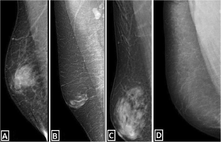
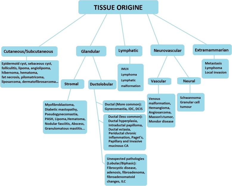
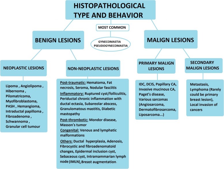
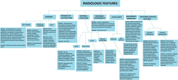
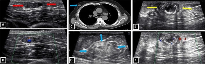
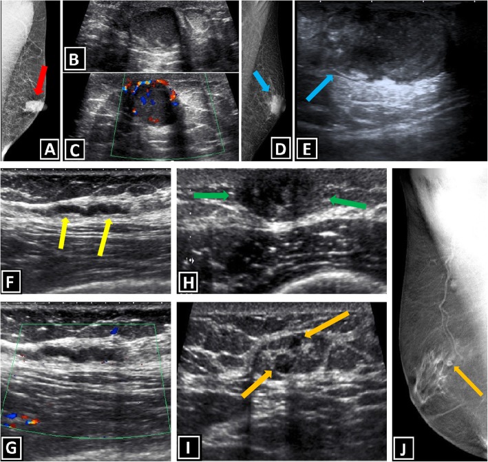
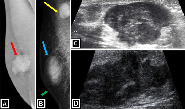
Male breast hosts various pathological conditions just like "female breast." However, histo-anatomical diversities with female breast lead to many differences regarding the frequency and presentation of diseases, the radiologic appearance of lesions, the diagnostic algorithm, and malignity features.Radiological modalities may play an important role in evaluating male breast lesions. Although some imaging findings are non-specific, having knowledge of certain imaging characteristics and radiologic patterns is the key to reduce the number of differential diagnoses or to reach an accurate diagnosis.Male breast imaging is mostly based on physical examination and is required for the complaints of palpable mass, breast enlargement, tenderness, nipple discharge, and nipple-skin changes. The majority of the male breast lumps are benign and the most common reason is gynecomastia. Although it is difficult to exclude malignancy in some cases, gynecomastia often has distinguishable imaging features. Pseudogynecomastia is another differential diagnosis that may be confused with gynecomastia. The distinction is important for the treatment plan.Apart from gynecomastia, other male breast lesions form a highly heterogeneous group and can be classified based on "Tissue origin," "Histopathological type and behavior," and "Radiologic features" for both simplification and comprehensive understanding.This article mainly focuses on emphasizing the results of basic histo-anatomical differences of male and female breasts, classifying male breast lesions, covering the spectrum of male breast diseases, and assisting radiologists in recognizing the imaging findings, in interpreting them through a holistic approach, in making a differential diagnosis, and in being a part of proper patient management.
男性乳房与“女性乳房”一样,会出现各种病理状况。然而,男性乳房与女性乳房在组织解剖学上的差异,导致了在疾病的发生率和表现、病变的放射学特征、诊断算法以及恶性特征等方面存在许多不同。放射学检查方法在评估男性乳房病变中可能起着重要作用。尽管一些影像学表现是非特异性的,但了解某些影像学特征和放射学模式是减少鉴别诊断数量或做出准确诊断的关键。男性乳房影像学检查主要基于体格检查,适用于可触及肿块、乳房增大、压痛、乳头溢液以及乳头 - 皮肤改变等症状的检查。大多数男性乳房肿块是良性的,最常见的原因是男性乳腺增生。虽然在某些情况下难以排除恶性病变,但男性乳腺增生通常具有可辨别的影像学特征。假性男性乳腺增生是另一种可能与男性乳腺增生相混淆的鉴别诊断。这种区分对于治疗方案很重要。除男性乳腺增生外,其他男性乳房病变构成了一个高度异质性的群体,可以根据“组织起源”“组织病理学类型和行为”以及“放射学特征”进行分类,以便简化和全面理解。本文主要着重强调男性和女性乳房基本组织解剖学差异的结果,对男性乳房病变进行分类,涵盖男性乳房疾病的范围,并协助放射科医生识别影像学表现,通过整体方法对其进行解读,做出鉴别诊断,并参与恰当的患者管理。