• 文献检索
  • 文档翻译
  • 深度研究
  • 学术资讯
  • Suppr Zotero 插件Zotero 插件
  • 邀请有礼
  • 套餐&价格
  • 历史记录
应用&插件
Suppr Zotero 插件Zotero 插件浏览器插件Mac 客户端Windows 客户端微信小程序
定价
高级版会员购买积分包购买API积分包
服务
文献检索文档翻译深度研究API 文档MCP 服务
关于我们
关于 Suppr公司介绍联系我们用户协议隐私条款
关注我们

Suppr 超能文献

核心技术专利:CN118964589B侵权必究
粤ICP备2023148730 号-1Suppr @ 2026

文献检索

告别复杂PubMed语法,用中文像聊天一样搜索,搜遍4000万医学文献。AI智能推荐,让科研检索更轻松。

立即免费搜索

文件翻译

保留排版,准确专业,支持PDF/Word/PPT等文件格式,支持 12+语言互译。

免费翻译文档

深度研究

AI帮你快速写综述,25分钟生成高质量综述,智能提取关键信息,辅助科研写作。

立即免费体验

自动定量提取和分析升主动脉中的 4D 流型:1.5T 和 3T 下的个体内比较。

Automated Quantitative Extraction and Analysis of 4D flow Patterns in the Ascending Aorta: An intraindividual comparison at 1.5 T and 3 T.

机构信息

Department of Diagnostic and Interventional Radiology, University of Leipzig - Heart Centre, Leipzig, Germany.

Department of Diagnostic and Interventional Radiology, University of Leipzig, Leipzig, Germany.

出版信息

Sci Rep. 2020 Feb 19;10(1):2949. doi: 10.1038/s41598-020-59826-2.

DOI:10.1038/s41598-020-59826-2
PMID:32076060
原文链接:https://pmc.ncbi.nlm.nih.gov/articles/PMC7031260/
Abstract

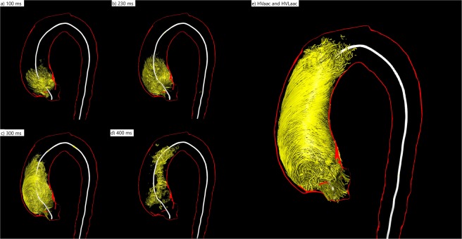
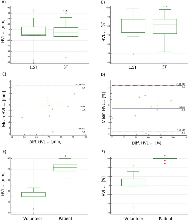
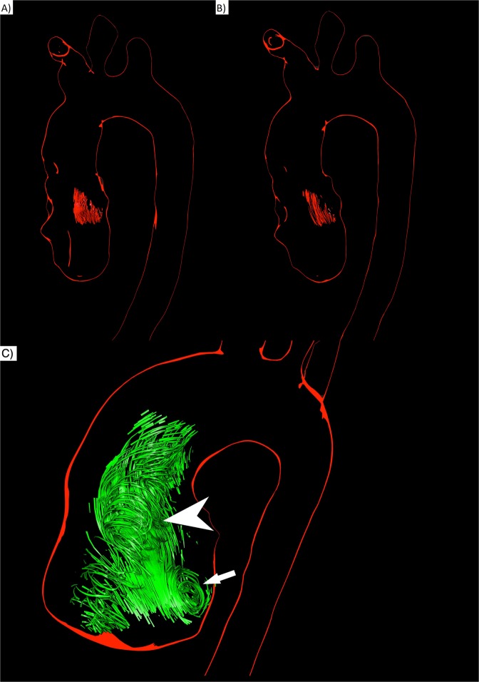
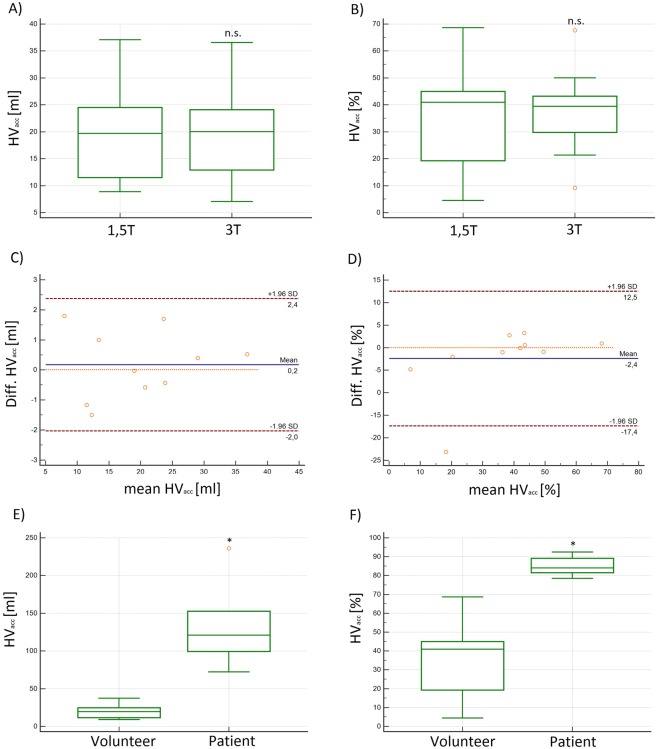
4D flow MRI enables quantitative assessment of helical flow. Current methods are susceptible to noise. To evaluate helical flow patterns in healthy volunteers and patients with bicuspid aortic valves (BAV) at 1.5 T and 3 T using pressure-based helix-extraction in 4D flow MRI. Two intraindividual 4D flow MRI examinations were performed at 1.5 T and 3 T in ten healthy volunteers (5 females, 32 ± 3 years) and 2 patients with BAV using different acceleration techniques (kt-GRAPPA and centra). Several new quantitative parameters for the evaluation of volumes [ml], lengths [mm] as well as temporal parameters [ms] of helical flow were introduced and analyzed using the software tool Bloodline. We found good correlations between measurements in volunteers at 1.5 T and 3 T regarding helical flow volumes (R = 0.98) and temporal existence (R = 0.99) of helices in the ascending aorta. Furthermore, we found significantly larger (11.7 vs. 77.6 ml) and longer lasting (317 vs. 769 ms) helices in patients with BAV than in volunteers. The assessed parameters do not depend on the magnetic field strength used for the acquisition. The technique of pressure-based extraction of 4D flow MRI pattern is suitable for differentiation of normal and pathological flow.

摘要

4D flow MRI 能够定量评估螺旋流。目前的方法容易受到噪声的影响。本研究旨在使用基于压力的 4D flow MRI 螺旋提取方法,在 1.5T 和 3T 下评估健康志愿者和二叶式主动脉瓣(BAV)患者的螺旋流动模式。在 1.5T 和 3T 下,使用不同的加速技术(kt-GRAPPA 和 centra)对 10 名健康志愿者(5 名女性,32±3 岁)和 2 名 BAV 患者进行了两次个体内 4D flow MRI 检查。使用 Bloodline 软件工具,引入了几个新的定量参数来评估螺旋流的体积[ml]、长度[mm]和时间参数[ms]。我们发现志愿者在 1.5T 和 3T 下的螺旋流体积(R=0.98)和升主动脉中螺旋存在的时间(R=0.99)的测量值之间存在良好的相关性。此外,我们发现 BAV 患者的螺旋流体积(11.7 vs. 77.6ml)和持续时间(317 vs. 769ms)明显大于志愿者。所评估的参数不依赖于采集时使用的磁场强度。基于压力的 4D flow MRI 模式提取技术适合于正常和病理血流的区分。

https://cdn.ncbi.nlm.nih.gov/pmc/blobs/778d/7031260/2ba2d6eee460/41598_2020_59826_Fig8_HTML.jpg
https://cdn.ncbi.nlm.nih.gov/pmc/blobs/778d/7031260/0165a99b89a6/41598_2020_59826_Fig1_HTML.jpg
https://cdn.ncbi.nlm.nih.gov/pmc/blobs/778d/7031260/a031c88f629f/41598_2020_59826_Fig2_HTML.jpg
https://cdn.ncbi.nlm.nih.gov/pmc/blobs/778d/7031260/bcf48c3370e1/41598_2020_59826_Fig3_HTML.jpg
https://cdn.ncbi.nlm.nih.gov/pmc/blobs/778d/7031260/a785c4ab4eed/41598_2020_59826_Fig4_HTML.jpg
https://cdn.ncbi.nlm.nih.gov/pmc/blobs/778d/7031260/d202919d2231/41598_2020_59826_Fig5_HTML.jpg
https://cdn.ncbi.nlm.nih.gov/pmc/blobs/778d/7031260/39b3434adf3d/41598_2020_59826_Fig6_HTML.jpg
https://cdn.ncbi.nlm.nih.gov/pmc/blobs/778d/7031260/541b0773ad40/41598_2020_59826_Fig7_HTML.jpg
https://cdn.ncbi.nlm.nih.gov/pmc/blobs/778d/7031260/2ba2d6eee460/41598_2020_59826_Fig8_HTML.jpg

相似文献

1
Automated Quantitative Extraction and Analysis of 4D flow Patterns in the Ascending Aorta: An intraindividual comparison at 1.5 T and 3 T.自动定量提取和分析升主动脉中的 4D 流型:1.5T 和 3T 下的个体内比较。
Sci Rep. 2020 Feb 19;10(1):2949. doi: 10.1038/s41598-020-59826-2.
2
Time-resolved 3-dimensional magnetic resonance phase contrast imaging (4D Flow MRI) reveals altered blood flow patterns in the ascending aorta of patients with valve-sparing aortic root replacement.时分辨 3 维磁共振相位对比成像(4DFlowMRI)显示,在保留主动脉瓣的主动脉根部置换术后患者的升主动脉中,血流模式发生改变。
J Thorac Cardiovasc Surg. 2020 Mar;159(3):798-810.e1. doi: 10.1016/j.jtcvs.2019.02.127. Epub 2019 Apr 5.
3
Wall shear stress and flow patterns in the ascending aorta in patients with bicuspid aortic valves differ significantly from tricuspid aortic valves: a prospective study.二叶式主动脉瓣患者升主动脉壁切应力和血流模式与三叶式主动脉瓣显著不同:一项前瞻性研究。
Eur Heart J Cardiovasc Imaging. 2013 Aug;14(8):797-804. doi: 10.1093/ehjci/jes273. Epub 2012 Dec 9.
4
Flow-sensitive four-dimensional magnetic resonance imaging: flow patterns in ascending aortic aneurysms.血流敏感型四维磁共振成像:升主动脉瘤中的血流模式
Eur J Cardiothorac Surg. 2008 Jul;34(1):11-6. doi: 10.1016/j.ejcts.2008.03.047. Epub 2008 Jun 2.
5
4D flow MRI, cardiac function, and T -mapping: Association of valve-mediated changes in aortic hemodynamics with left ventricular remodeling.4D 血流 MRI、心功能和 T 映射:主动脉血流动力学中瓣膜介导的变化与左心室重构的关系。
J Magn Reson Imaging. 2018 Jul;48(1):121-131. doi: 10.1002/jmri.25916. Epub 2017 Dec 5.
6
k-t accelerated aortic 4D flow MRI in under two minutes: Feasibility and impact of resolution, k-space sampling patterns, and respiratory navigator gating on hemodynamic measurements.在两分钟内完成 k-t 加速主动脉 4D 流 MRI:分辨率、k 空间采样模式和呼吸导航门控对血流测量的可行性和影响。
Magn Reson Med. 2018 Jan;79(1):195-207. doi: 10.1002/mrm.26661. Epub 2017 Mar 7.
7
Impact of sequence type and field strength (1.5, 3, and 7T) on 4D flow MRI hemodynamic aortic parameters in healthy volunteers.健康志愿者中序列类型和磁场强度(1.5T、3T 和 7T)对 4D 流 MRI 血流动力学主动脉参数的影响。
Magn Reson Med. 2021 Feb;85(2):721-733. doi: 10.1002/mrm.28450. Epub 2020 Aug 4.
8
Noninvasive 4D pressure difference mapping derived from 4D flow MRI in patients with repaired aortic coarctation: comparison with young healthy volunteers.无创4D压差映射技术源自修复性主动脉缩窄患者的4D流动MRI:与年轻健康志愿者的比较。
Int J Cardiovasc Imaging. 2015 Apr;31(4):823-30. doi: 10.1007/s10554-015-0604-3. Epub 2015 Feb 3.
9
Quantitative Analysis of Vortical Blood Flow in the Thoracic Aorta Using 4D Phase Contrast MRI.使用四维相位对比磁共振成像对胸主动脉内的涡流进行定量分析。
PLoS One. 2015 Sep 29;10(9):e0139025. doi: 10.1371/journal.pone.0139025. eCollection 2015.
10
Bileaflet mechanical aortic valves do not alter ascending aortic wall shear stress.双叶机械主动脉瓣不会改变升主动脉壁剪切应力。
Int J Cardiovasc Imaging. 2019 Apr;35(4):703-710. doi: 10.1007/s10554-018-1508-9. Epub 2019 Feb 11.

引用本文的文献

1
Validation of 2D flow MRI for helical and vortical flows.二维流磁共振成像在螺旋和涡旋流中的验证。
Open Heart. 2024 Mar 8;11(1):e002451. doi: 10.1136/openhrt-2023-002451.
2
Comparison of aortic blood flow rotational direction in healthy volunteers and patients with bicuspid aortic valves using volumetric velocity-sensitive cardiovascular magnetic resonance imaging.使用容积速度敏感型心血管磁共振成像比较健康志愿者和二叶式主动脉瓣患者的主动脉血流旋转方向。
Quant Imaging Med Surg. 2023 Dec 1;13(12):7973-7986. doi: 10.21037/qims-23-183. Epub 2023 Oct 27.
3
Restoration of flow in the aorta: a novel therapeutic target in aortic valve intervention.

本文引用的文献

1
Intracardiac 4D Flow MRI in Congenital Heart Disease: Recommendations on Behalf of the ISMRM Flow & Motion Study Group.先天性心脏病的心脏内 4D 流 MRI:ISMRM 流动与运动研究组的建议。
J Magn Reson Imaging. 2019 Sep;50(3):677-681. doi: 10.1002/jmri.26858. Epub 2019 Jul 17.
2
Aortic dilatation associates with flow displacement and increased circumferential wall shear stress in patients without aortic stenosis: A prospective clinical study.在没有主动脉瓣狭窄的患者中,主动脉扩张与血流移位和周向壁切应力增加相关:一项前瞻性临床研究。
J Magn Reson Imaging. 2019 Jul;50(1):136-145. doi: 10.1002/jmri.26655. Epub 2019 Jan 18.
3
恢复主动脉血流:主动脉瓣介入治疗的新靶点。
Nat Rev Cardiol. 2024 Apr;21(4):264-273. doi: 10.1038/s41569-023-00943-6. Epub 2023 Oct 25.
4
Possible Early Generation of Physiological Helical Flow Could Benefit the Triflo Trileaflet Heart Valve Prosthesis Compared to Bileaflet Valves.与双叶瓣心脏瓣膜相比,生理性螺旋血流的早期形成可能使三叶瓣Triflo心脏瓣膜假体受益。
Bioengineering (Basel). 2020 Dec 8;7(4):158. doi: 10.3390/bioengineering7040158.
Age-related changes of shape and flow dynamics in healthy adult aortas: A 4D flow MRI study.
健康成年人升主动脉形态和流动力学的年龄相关性变化:4D 血流 MRI 研究。
J Magn Reson Imaging. 2019 Jan;49(1):90-100. doi: 10.1002/jmri.26210. Epub 2018 Aug 13.
4
Advanced Analysis Techniques for Intra-cardiac Flow Evaluation from 4D Flow MRI.基于4D流磁共振成像的心脏内血流评估的先进分析技术
Curr Radiol Rep. 2016;4:38. doi: 10.1007/s40134-016-0167-7. Epub 2016 May 20.
5
Quantitative Analysis of Vortical Blood Flow in the Thoracic Aorta Using 4D Phase Contrast MRI.使用四维相位对比磁共振成像对胸主动脉内的涡流进行定量分析。
PLoS One. 2015 Sep 29;10(9):e0139025. doi: 10.1371/journal.pone.0139025. eCollection 2015.
6
4D flow cardiovascular magnetic resonance consensus statement.4D 流动心血管磁共振共识声明。
J Cardiovasc Magn Reson. 2015 Aug 10;17(1):72. doi: 10.1186/s12968-015-0174-5.
7
4D flow imaging with MRI.利用磁共振成像进行四维血流成像。
Cardiovasc Diagn Ther. 2014 Apr;4(2):173-92. doi: 10.3978/j.issn.2223-3652.2014.01.02.
8
Aortic dilation in bicuspid aortic valve disease: flow pattern is a major contributor and differs with valve fusion type.二叶式主动脉瓣病变中的主动脉扩张:血流模式是主要贡献因素,且其随瓣叶融合类型而异。
Circ Cardiovasc Imaging. 2013 Jul;6(4):499-507. doi: 10.1161/CIRCIMAGING.113.000528. Epub 2013 Jun 14.
9
4D flow MRI.四维磁共振血流成像技术。
J Magn Reson Imaging. 2012 Nov;36(5):1015-36. doi: 10.1002/jmri.23632.
10
Flow-sensitive 4D MRI of the thoracic aorta: comparison of image quality, quantitative flow, and wall parameters at 1.5 T and 3 T.胸主动脉的流量敏感 4D MRI:1.5T 和 3T 时图像质量、定量流量和管壁参数的比较。
J Magn Reson Imaging. 2012 Nov;36(5):1097-103. doi: 10.1002/jmri.23735. Epub 2012 Jun 28.