Tan Jiaxiong, Yu Zhi, Huang Jingying, Chen Youchun, Huang Shuxin, Yao Danlin, Xu Ling, Lu Yuhong, Chen Shaohua, Li Yangqiu
1Department of Hematology, First Affiliated Hospital, Jinan University, Guangzhou, 510632 China.
2Institute of Hematology, School of Medicine, Key Laboratory for Regenerative Medicine of Ministry of Education, Jinan University, Guangzhou, 510632 China.
Biomark Res. 2020 Feb 13;8:6. doi: 10.1186/s40364-020-0185-8. eCollection 2020.
Altered expression of T cell immune inhibitory receptors may result in immunosuppression and associate with the poor prognosis of leukemia patients in which the leukemic bone marrow (BM) microenvironment may contribute to such immunosuppression. We found higher numbers of programmed death-1 (PD-1) + exhausted T cells in peripheral blood (PB) from acute myeloid leukemia (AML) patients. To investigate the leukemic BM influence on immunosuppression, we further compared the distributions of PD-1 and T cell immunoglobulin mucin-3 (Tim-3) and the exhausted T cell phenotype in PB and BM from AML patients and characterized their relationship with clinical outcome.
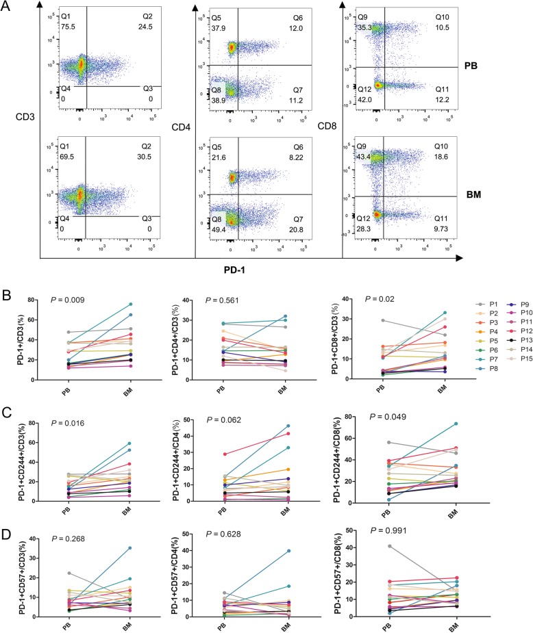
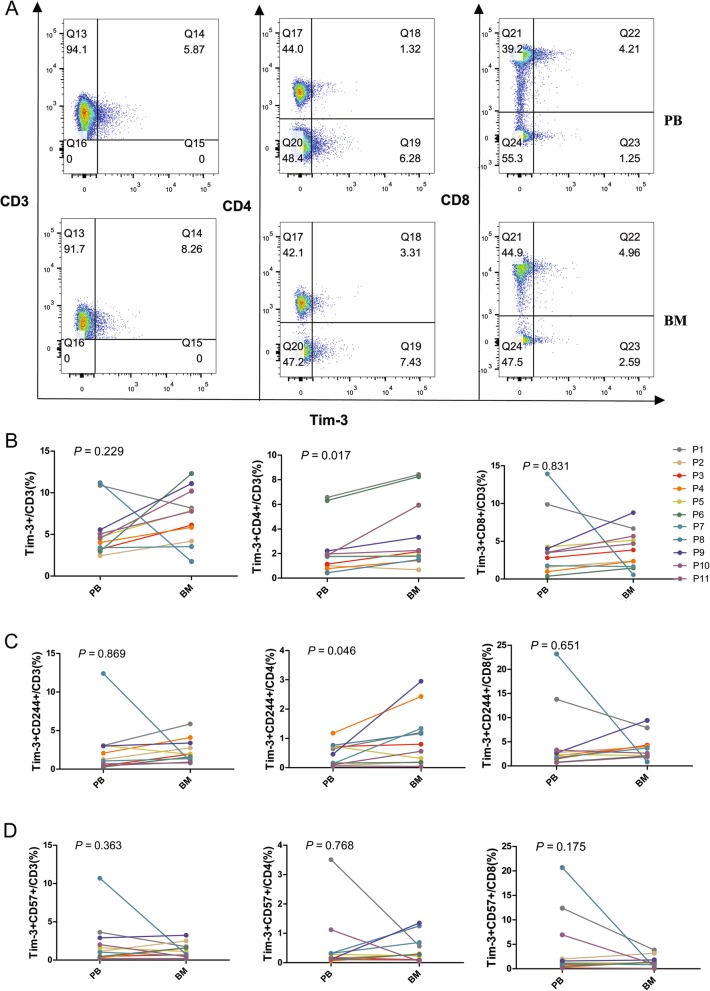
PB and BM samples from 15 patients with newly diagnosed AML were collected and analyzed for the expression of PD-1, Tim-3, CD244, and CD57 on CD3+, CD4+, and CD8+ T cells by multicolor flow cytometry.
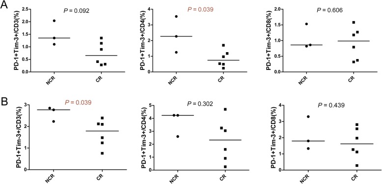
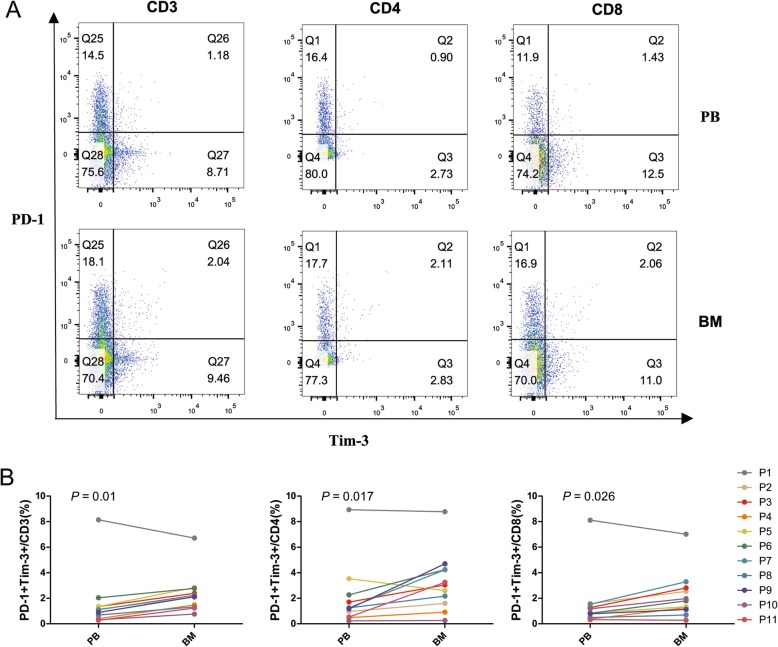
The proportions of PD-1 + CD3+ and PD-1 + CD8+ T cells were significantly higher in BM compared with PB. Similarly, higher PD-1 CD244 CD3 and PD-1 CD244 CD8 T cells were found in BM, and an increased tendency for PD-1 + CD244 + CD4+ T cells was also detected in this group. In contrast, increased Tim-3 + CD4+/Tim-3 + CD244 + CD4+ T cells were predominant in BM compared with PB, but there was no statistically significant difference in Tim-3 + CD8+ T cells. Moreover, PD-1 and Tim-3 double-positive CD3+/CD4+/CD8+ T cells were significantly increased in the BM group. In addition, a higher proportion of PD-1 + Tim-3 + CD3+ T cells in the BM and PD-1 + Tim-3 + CD4+ T cells in PB was detected in non-complete remission (NCR) compared with complete remission (CR) patients after first-cycle chemotherapy.
Upregulation of PD-1 and Tim-3 and the exhausted phenotype of CD4+ and CD8+ T cells in the BM of AML patients may contribute to mediating the leukemic immunosuppressive microenvironment, and increased PD-1 + Tim-3+ CD8+ T cells may be related to T cell dysfunction in AML, which may influence clinical outcome.
T细胞免疫抑制受体表达改变可能导致免疫抑制,并与白血病患者的不良预后相关,其中白血病骨髓(BM)微环境可能促成这种免疫抑制。我们发现急性髓系白血病(AML)患者外周血(PB)中程序性死亡1(PD-1)+耗竭性T细胞数量更多。为了研究白血病BM对免疫抑制的影响,我们进一步比较了AML患者PB和BM中PD-1和T细胞免疫球蛋白粘蛋白3(Tim-3)的分布以及耗竭性T细胞表型,并确定它们与临床结局的关系。
收集15例新诊断AML患者的PB和BM样本,通过多色流式细胞术分析CD3+、CD4+和CD8+ T细胞上PD-1、Tim-3、CD244和CD57的表达。
与PB相比,BM中PD-1+CD3+和PD-1+CD8+ T细胞的比例显著更高。同样,BM中发现更高比例的PD-1 CD244 CD3和PD-1 CD244 CD8 T细胞,并且在该组中也检测到PD-1+CD244+CD4+ T细胞有增加趋势。相比之下,与PB相比,BM中Tim-3+CD4+/Tim-3+CD244+CD4+ T细胞增加更为明显,但Tim-3+CD8+ T细胞无统计学显著差异。此外,BM组中PD-1和Tim-3双阳性CD3+/CD4+/CD8+ T细胞显著增加。另外,与首次化疗后完全缓解(CR)患者相比,非完全缓解(NCR)患者BM中PD-1+Tim-3+CD3+ T细胞比例更高,PB中PD-1+Tim-3+CD4+ T细胞比例更高。
AML患者BM中PD-1和Tim-3上调以及CD4+和CD8+ T细胞的耗竭表型可能有助于介导白血病免疫抑制微环境,并且PD-1+Tim-3+CD8+ T细胞增加可能与AML中T细胞功能障碍有关,这可能影响临床结局。