• 文献检索
  • 文档翻译
  • 深度研究
  • 学术资讯
  • Suppr Zotero 插件Zotero 插件
  • 邀请有礼
  • 套餐&价格
  • 历史记录
应用&插件
Suppr Zotero 插件Zotero 插件浏览器插件Mac 客户端Windows 客户端微信小程序
定价
高级版会员购买积分包购买API积分包
服务
文献检索文档翻译深度研究API 文档MCP 服务
关于我们
关于 Suppr公司介绍联系我们用户协议隐私条款
关注我们

Suppr 超能文献

核心技术专利:CN118964589B侵权必究
粤ICP备2023148730 号-1Suppr @ 2026

文献检索

告别复杂PubMed语法,用中文像聊天一样搜索,搜遍4000万医学文献。AI智能推荐,让科研检索更轻松。

立即免费搜索

文件翻译

保留排版,准确专业,支持PDF/Word/PPT等文件格式,支持 12+语言互译。

免费翻译文档

深度研究

AI帮你快速写综述,25分钟生成高质量综述,智能提取关键信息,辅助科研写作。

立即免费体验

下胫腓联合复位不良可能降低固定稳定性:一项生物力学研究。

Syndesmotic malreduction may decrease fixation stability: a biomechanical study.

机构信息

Department of Sports Medicine, Peking University Shenzhen Hospital, #1120 Lianhua Road, Shenzhen, Guangdong Province, China.

National and Local Joint Engineering Research Center of Orthopaedic Biomaterials, Peking University Shenzhen Hospital, #1120 Lianhua Road, Shenzhen, Guangdong Province, China.

出版信息

J Orthop Surg Res. 2020 Feb 21;15(1):64. doi: 10.1186/s13018-020-01584-y.

DOI:10.1186/s13018-020-01584-y
PMID:32085779
原文链接:https://pmc.ncbi.nlm.nih.gov/articles/PMC7035663/
Abstract

BACKGROUND

This study aims to investigate the malreduction of syndesmosis and its effects on stability.

METHODS

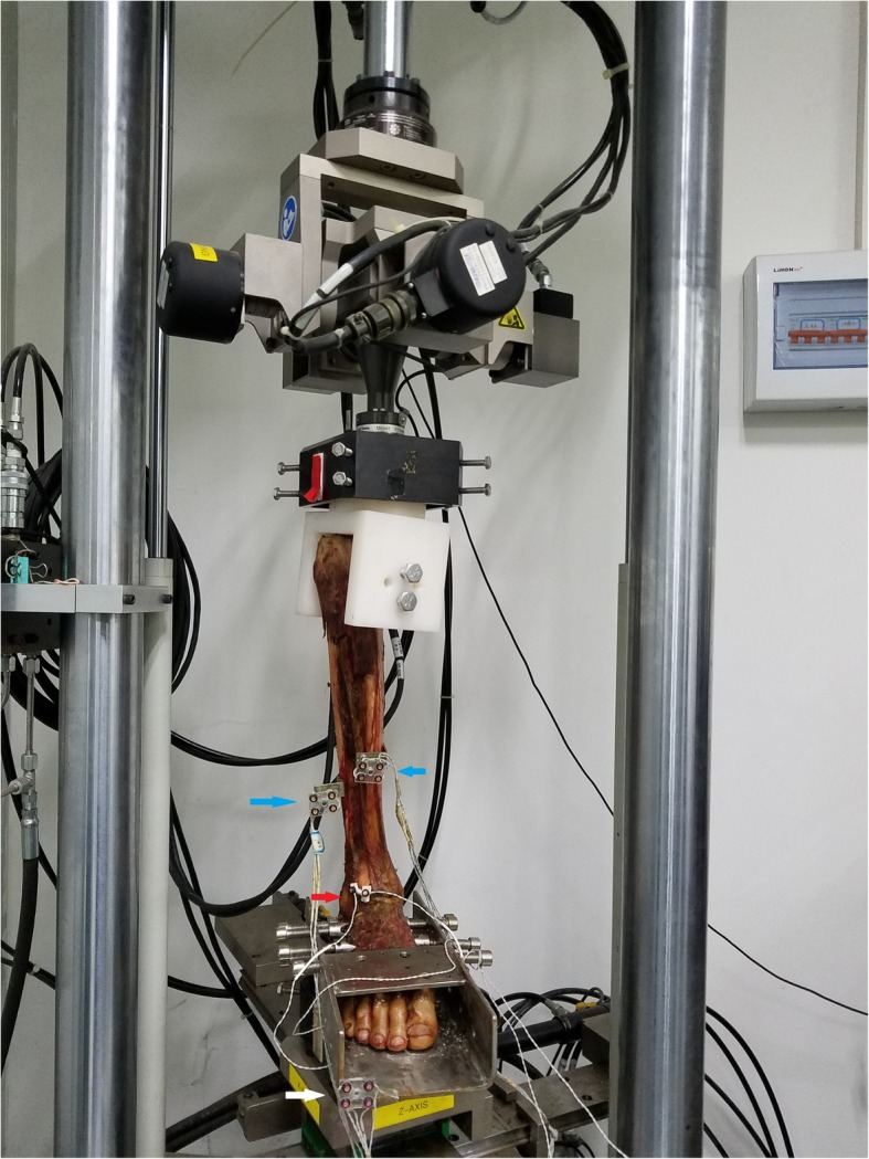
The biomechanical tests, including the three-dimensional (3D) displacement of the syndesmotic incisura, fibular rotation angle, and torque resistance, were performed on six cadaver legs. These specimens were first tested intact (intact group), then cut all the syndesmotic ligaments and fixed in anatomical position (anatomical model group) and test again. After that, syndesmosis was fixed in 1 cm malreduction (anterior and posterior displacement group) to do the same test.

RESULTS

In internal or external load, there were significant differences in torque resistance and fibular rotation angle (internal t = 2.412, P = 0.036; external t = 2.412, P = 0.039) between the intact and post-malreduction groups. In internal rotation load, there were significant differences in sagittal displacement between the intact and post-malreduction groups (P = 0.011), and between the anatomical and post-malreduction groups (P = 0.020). In external rotation load, significant differences existed between the intact and ant-malreduction group (P = 0.034) in sagittal (anterior-posterior) displacement. Significant differences also existed between the intact and post-malreduction groups (P = 0.013), and between the anatomical and post-malreduction groups (P = 0.038) in coronal (medial-lateral) displacement.

CONCLUSIONS

Malreduction in different conditions does affect the stability of the syndesmotic fixation. The result of the study may reveal the biomechanical mechanism of poor clinical outcome in syndesmosis malreduction patients and pathological displacement patterns of the ankle under syndesmotic malreduction conditions.

LEVEL OF EVIDENCE

III.

摘要

背景

本研究旨在探讨下胫腓联合复位不良及其对稳定性的影响。

方法

对 6 例尸体下肢进行生物力学测试,包括下胫腓联合切迹的三维(3D)位移、腓骨旋转角度和抗扭阻力。这些标本首先进行完整测试(完整组),然后切断所有下胫腓联合韧带并在解剖位置固定(解剖模型组),再进行测试。之后,将下胫腓联合固定在 1cm 复位不良(前后移位组)进行相同的测试。

结果

在内外负荷下,完整组和复位后组之间的抗扭阻力和腓骨旋转角度存在显著差异(内旋 t=2.412,P=0.036;外旋 t=2.412,P=0.039)。在旋转负荷下,完整组和复位后组之间在矢状面位移上存在显著差异(P=0.011),解剖模型组和复位后组之间也存在显著差异(P=0.020)。在外旋负荷下,完整组和前复位组在矢状面(前后)位移上存在显著差异(P=0.034)。完整组和复位后组之间以及解剖模型组和复位后组之间在冠状面(内外)位移上也存在显著差异(P=0.013,P=0.038)。

结论

不同条件下的复位不良确实会影响下胫腓联合固定的稳定性。研究结果可能揭示了下胫腓联合复位不良患者临床结果不佳的生物力学机制以及下胫腓联合复位不良情况下踝关节的病理性移位模式。

证据水平

III。

https://cdn.ncbi.nlm.nih.gov/pmc/blobs/3dc8/7035663/8de9edc530bf/13018_2020_1584_Fig4_HTML.jpg
https://cdn.ncbi.nlm.nih.gov/pmc/blobs/3dc8/7035663/b0d63088ae28/13018_2020_1584_Fig1_HTML.jpg
https://cdn.ncbi.nlm.nih.gov/pmc/blobs/3dc8/7035663/e3ecf15e4f17/13018_2020_1584_Fig2_HTML.jpg
https://cdn.ncbi.nlm.nih.gov/pmc/blobs/3dc8/7035663/e63e8509f8f4/13018_2020_1584_Fig3_HTML.jpg
https://cdn.ncbi.nlm.nih.gov/pmc/blobs/3dc8/7035663/8de9edc530bf/13018_2020_1584_Fig4_HTML.jpg

相似文献

1
Syndesmotic malreduction may decrease fixation stability: a biomechanical study.下胫腓联合复位不良可能降低固定稳定性:一项生物力学研究。
J Orthop Surg Res. 2020 Feb 21;15(1):64. doi: 10.1186/s13018-020-01584-y.
2
Overtightening of the syndesmosis revisited and the effect of syndesmotic malreduction on ankle dorsiflexion.下胫腓联合过度收紧再探讨及下胫腓联合复位不良对踝关节背屈的影响
Injury. 2017 Jun;48(6):1253-1257. doi: 10.1016/j.injury.2017.03.029. Epub 2017 Mar 25.
3
Effect of Posterior Malleolus Fracture on Syndesmotic Reduction: A Cadaveric Study.后踝骨折对下胫腓联合复位的影响:一项尸体研究。
J Bone Joint Surg Am. 2018 Feb 7;100(3):243-248. doi: 10.2106/JBJS.17.00217.
4
Novel anatomical reconstruction of distal tibiofibular ligaments restores syndesmotic biomechanics.胫腓骨远端韧带的新型解剖重建可恢复下胫腓联合的生物力学。
Knee Surg Sports Traumatol Arthrosc. 2017 Jun;25(6):1866-1872. doi: 10.1007/s00167-017-4485-y. Epub 2017 Mar 20.
5
Correlation of Incisura Anatomy With Syndesmotic Malreduction.切迹解剖与下胫腓联合复位不良的相关性
Foot Ankle Int. 2018 Mar;39(3):369-375. doi: 10.1177/1071100717744332. Epub 2017 Dec 18.
6
Assessment of malreduction standards for the syndesmosis in bilateral CT scans of uninjured ankles.双侧 CT 扫描未受伤踝关节下胫腓联合复位不良的评估标准。
Bone Joint J. 2021 Jan;103-B(1):178-183. doi: 10.1302/0301-620X.103B1.BJJ-2020-0844.R1.
7
Medial Clamp Tine Positioning Affects Ankle Syndesmosis Malreduction.内侧夹齿位置影响踝关节下胫腓联合复位不良。
J Orthop Trauma. 2017 Aug;31(8):440-446. doi: 10.1097/BOT.0000000000000882.
8
Arthroscopically measured syndesmotic stability after screw vs. suture button fixation in a cadaveric model.在尸体模型中,对比螺钉与缝线纽扣固定后关节镜测量下的下胫腓联合稳定性。
Injury. 2017 Nov;48(11):2433-2437. doi: 10.1016/j.injury.2017.08.066. Epub 2017 Aug 31.
9
The effect of suture-button fixation on simulated syndesmotic malreduction: a cadaveric study.缝线纽扣固定对模拟下胫腓联合复位不良的影响:一项尸体研究。
J Bone Joint Surg Am. 2014 Oct 15;96(20):1732-8. doi: 10.2106/JBJS.N.00198.
10
Incisura Morphology as a Risk Factor for Syndesmotic Malreduction.切迹形态作为下胫腓联合复位不良的危险因素
Foot Ankle Int. 2016 Jul;37(7):748-54. doi: 10.1177/1071100716637709. Epub 2016 Mar 15.

引用本文的文献

1
Anatomy, biomechanics, and clinical advances of proximal tibiofibular joint in pain management.近端胫腓关节在疼痛管理中的解剖学、生物力学及临床进展
Front Surg. 2025 Jul 17;12:1604538. doi: 10.3389/fsurg.2025.1604538. eCollection 2025.
2
Results of open reduction and internal fixation of proximal humerus fractures using a proximal humeral plating system with smooth pegs.使用带有光滑钉的肱骨近端接骨板系统对肱骨近端骨折进行切开复位内固定的结果
Shoulder Elbow. 2025 Jan 15:17585732241309582. doi: 10.1177/17585732241309582.
3
Residual coronary malformation after tibial shaft fracture alters the contact status of the meniscus and cartilage in the knee joint: a computational study.

本文引用的文献

1
Incidence and severity of malreduction of the tibiofibular syndesmosis following surgical treatement of displaced ankle fractures and impact on the function -Clinical study and MRI evaluation.踝关节移位骨折手术治疗后胫腓下联合复位不良的发生率、严重程度及其对功能的影响——临床研究与MRI评估
Injury. 2018 Jun;49(6):1220-1227. doi: 10.1016/j.injury.2018.04.027. Epub 2018 Apr 23.
2
Is It Possible to Overcompress the Syndesmosis?下胫腓联合有可能过度压缩吗?
J Foot Ankle Surg. 2018 Sep-Oct;57(5):1005-1009. doi: 10.1053/j.jfas.2017.11.037. Epub 2018 Mar 13.
3
Distal Tibiofibular Syndesmosis: Anatomy, Biomechanics, Injury and Management.
胫骨干骨折后残留的冠状动脉畸形改变膝关节半月板和软骨的接触状态:一项计算研究。
Front Surg. 2024 Sep 13;11:1325085. doi: 10.3389/fsurg.2024.1325085. eCollection 2024.
4
Comparison of Two Different Methods to Evaluate Ankle Syndesmosis on Lateral Ankle Radiographs.两种不同方法在踝关节侧位X线片上评估下胫腓联合的比较。
Cureus. 2023 Dec 25;15(12):e51070. doi: 10.7759/cureus.51070. eCollection 2023 Dec.
5
Maisonneuve fracture treated with suture-button system stabilization combined with plate and arthroscopic assistance (SBPAA): Clinical and radiological evaluation in short-medium period.采用缝线纽扣系统固定联合钢板及关节镜辅助治疗(SBPAA)的马松纽夫骨折:短期至中期的临床及影像学评估
J Orthop. 2023 Oct 14;46:12-17. doi: 10.1016/j.jor.2023.10.007. eCollection 2023 Dec.
6
"Flexible nature of fixation" in syndesmotic stabilization of the inferior tibiofibular joint affects the radiological reduction outcome.下胫腓联合联合固定的“固定灵活性”影响放射学复位结果。
Int Orthop. 2022 Nov;46(11):2649-2657. doi: 10.1007/s00264-022-05550-7. Epub 2022 Aug 19.
7
Intraoperative Assessment of Reduction of the Ankle Syndesmosis.踝关节下胫腓联合复位的术中评估
Curr Rev Musculoskelet Med. 2022 Oct;15(5):344-352. doi: 10.1007/s12178-022-09769-0. Epub 2022 Jul 13.
8
A 10-Year Follow-Up of Ankle Syndesmotic Injuries: Prospective Comparison of Knotless Suture-Button Fixation and Syndesmotic Screw Fixation.踝关节下胫腓联合损伤的10年随访:无结缝线纽扣固定与下胫腓联合螺钉固定的前瞻性比较
J Clin Med. 2022 Apr 30;11(9):2524. doi: 10.3390/jcm11092524.
9
Evidence-Based Surgical Treatment Algorithm for Unstable Syndesmotic Injuries.不稳定下胫腓联合损伤的循证外科治疗算法
J Clin Med. 2022 Jan 10;11(2):331. doi: 10.3390/jcm11020331.
胫腓下联合:解剖、生物力学、损伤与治疗
Open Orthop J. 2017 Jul 31;11:670-677. doi: 10.2174/1874325001711010670. eCollection 2017.
4
Syndesmotic Ankle Fractures.下胫腓联合踝关节骨折
J Orthop Trauma. 2018 Jan;32(1):10-14. doi: 10.1097/BOT.0000000000000937.
5
Novel anatomical reconstruction of distal tibiofibular ligaments restores syndesmotic biomechanics.胫腓骨远端韧带的新型解剖重建可恢复下胫腓联合的生物力学。
Knee Surg Sports Traumatol Arthrosc. 2017 Jun;25(6):1866-1872. doi: 10.1007/s00167-017-4485-y. Epub 2017 Mar 20.
6
Effect of Complete Syndesmotic Disruption and Deltoid Injuries and Different Reduction Methods on Ankle Joint Contact Mechanics.下胫腓联合完全断裂、三角韧带损伤及不同复位方法对踝关节接触力学的影响
Foot Ankle Int. 2017 Jun;38(6):694-700. doi: 10.1177/1071100717696360. Epub 2017 Mar 15.
7
An Update on Management of Syndesmosis Injury: A National US Database Study.下胫腓联合损伤治疗的最新进展:一项美国全国性数据库研究
Am J Orthop (Belle Mead NJ). 2016 Nov/Dec;45(7):E472-E477.
8
Three-Dimensional Analysis of Fibular Motion After Fixation of Syndesmotic Injuries With a Screw or Suture-Button Construct.使用螺钉或缝线纽扣结构固定下胫腓联合损伤后腓骨运动的三维分析
Foot Ankle Int. 2016 Dec;37(12):1350-1356. doi: 10.1177/1071100716666865. Epub 2016 Sep 20.
9
A Comparison of Screw Fixation and Suture-Button Fixation in a Syndesmosis Injury in an Ankle Fracture.踝关节骨折下胫腓联合损伤中螺钉固定与缝线纽扣固定的比较
J Foot Ankle Surg. 2016 Sep-Oct;55(5):985-90. doi: 10.1053/j.jfas.2016.05.002. Epub 2016 Jul 19.
10
Three-dimensional motions of distal syndesmosis during walking.行走过程中胫腓下联合的三维运动
J Orthop Surg Res. 2015 Oct 24;10:166. doi: 10.1186/s13018-015-0306-5.