• 文献检索
  • 文档翻译
  • 深度研究
  • 学术资讯
  • Suppr Zotero 插件Zotero 插件
  • 邀请有礼
  • 套餐&价格
  • 历史记录
应用&插件
Suppr Zotero 插件Zotero 插件浏览器插件Mac 客户端Windows 客户端微信小程序
定价
高级版会员购买积分包购买API积分包
服务
文献检索文档翻译深度研究API 文档MCP 服务
关于我们
关于 Suppr公司介绍联系我们用户协议隐私条款
关注我们

Suppr 超能文献

核心技术专利:CN118964589B侵权必究
粤ICP备2023148730 号-1Suppr @ 2026

文献检索

告别复杂PubMed语法,用中文像聊天一样搜索,搜遍4000万医学文献。AI智能推荐,让科研检索更轻松。

立即免费搜索

文件翻译

保留排版,准确专业,支持PDF/Word/PPT等文件格式,支持 12+语言互译。

免费翻译文档

深度研究

AI帮你快速写综述,25分钟生成高质量综述,智能提取关键信息,辅助科研写作。

立即免费体验

角膜共焦显微镜检测痛性糖尿病周围神经病患者的小纤维神经损伤。

Corneal confocal microscopy detects small nerve fibre damage in patients with painful diabetic neuropathy.

机构信息

Division of Cardiovascular Sciences, Cardiac Centre, Faculty of Medical and Human Sciences, University of Manchester and NIHR/Wellcome Trust Clinical Research Facility, Manchester, UK.

Cardiovascular Trials Unit, Central Manchester University Hospital NHS Foundation Trust, Manchester, UK.

出版信息

Sci Rep. 2020 Feb 25;10(1):3371. doi: 10.1038/s41598-020-60422-7.

DOI:10.1038/s41598-020-60422-7
PMID:32099076
原文链接:https://pmc.ncbi.nlm.nih.gov/articles/PMC7042367/
Abstract

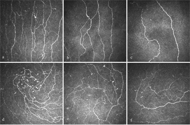
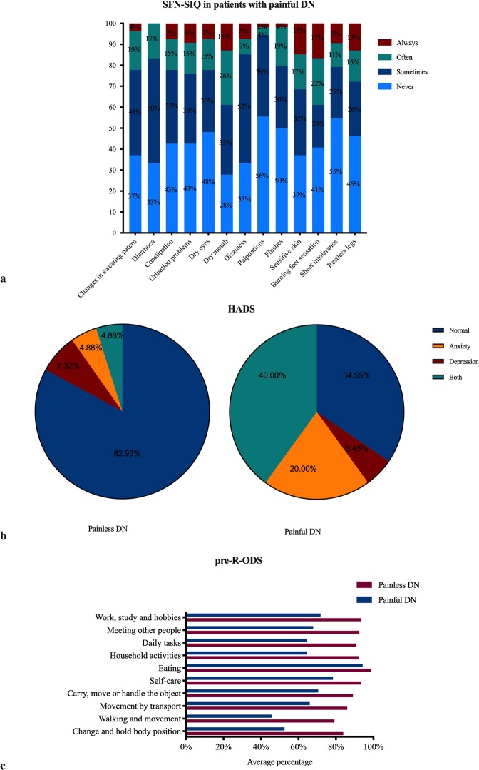
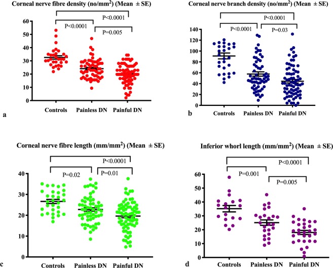
Neuropathic pain is believed to arise from damage to nociceptive C fibres in diabetic neuropathy (DN). We have utilised corneal confocal microscopy (CCM) to quantify the severity of small nerve fibre damage in relation to the severity of neuropathic pain and quality of life (QoL) in patients with and without painful DN. 30 controls and patients with painful (n = 78) and painless (n = 62) DN underwent assessment of large and small nerve fibre function, CCM, neuropathic symptoms (small fibre neuropathy symptom inventory questionnaire, neuropathic pain scale) and QoL (SF-36, pre-R-ODS and hospital anxiety and depression scale). Patients with painful compared to painless DN, had comparable neurophysiology and vibration perception, but lower corneal nerve fibre density (20.1 ± 0.87 vs. 24.13 ± 0.91, P = 0.005), branch density (44.4 ± 3.31 vs. 57.74 ± 3.98, P = 0.03), length (19.61 ± 0.81 vs. 22.77 ± 0.83, P = 0.01), inferior whorl length (18.03 ± 1.46 vs. 25.1 ± 1.95, P = 0.005) and cold sensation threshold (21.35 ± 0.99 vs. 26.08 ± 0.5, P < 0.0001) and higher warm sensation threshold (43.7 ± 0.49 vs. 41.37 ± 0.51, P = 0.004) indicative of small fibre damage. There was a significant association between all CCM parameters and the severity of painful neuropathic symptoms, depression score and QoL. CCM identifies small nerve fibre loss, which correlates with the severity of neuropathic symptoms and reduced QoL in patients with painful diabetic neuropathy.

摘要

神经病理性疼痛被认为源于糖尿病性周围神经病(DN)中伤害感受 C 纤维的损伤。我们利用角膜共聚焦显微镜(CCM)来量化小纤维损伤的严重程度与有或无疼痛性 DN 的患者的神经病理性疼痛严重程度和生活质量(QoL)之间的关系。30 名对照者和有疼痛性(n=78)和无痛性(n=62)DN 的患者接受了大纤维和小纤维功能、CCM、神经病理性症状(小纤维神经病症状量表问卷、神经病理性疼痛量表)和 QoL(SF-36、预 R-ODS 和医院焦虑和抑郁量表)评估。与无痛性 DN 相比,疼痛性 DN 的患者具有可比的神经生理学和振动感觉,但角膜神经纤维密度更低(20.1±0.87 对 24.13±0.91,P=0.005)、分支密度更低(44.4±3.31 对 57.74±3.98,P=0.03)、长度更短(19.61±0.81 对 22.77±0.83,P=0.01)、下涡旋长度更短(18.03±1.46 对 25.1±1.95,P=0.005)和冷感觉阈值更高(21.35±0.99 对 26.08±0.5,P<0.0001)以及温觉阈值更高(43.7±0.49 对 41.37±0.51,P=0.004),提示小纤维损伤。所有 CCM 参数与疼痛性神经病理性症状的严重程度、抑郁评分和 QoL 之间均存在显著相关性。CCM 可识别小神经纤维的丧失,这与疼痛性糖尿病性周围神经病患者的神经病理性症状严重程度和 QoL 降低相关。

https://cdn.ncbi.nlm.nih.gov/pmc/blobs/8595/7042367/0d60da823439/41598_2020_60422_Fig3_HTML.jpg
https://cdn.ncbi.nlm.nih.gov/pmc/blobs/8595/7042367/56d5a6e3beb5/41598_2020_60422_Fig1_HTML.jpg
https://cdn.ncbi.nlm.nih.gov/pmc/blobs/8595/7042367/5d80dec9e543/41598_2020_60422_Fig2_HTML.jpg
https://cdn.ncbi.nlm.nih.gov/pmc/blobs/8595/7042367/0d60da823439/41598_2020_60422_Fig3_HTML.jpg
https://cdn.ncbi.nlm.nih.gov/pmc/blobs/8595/7042367/56d5a6e3beb5/41598_2020_60422_Fig1_HTML.jpg
https://cdn.ncbi.nlm.nih.gov/pmc/blobs/8595/7042367/5d80dec9e543/41598_2020_60422_Fig2_HTML.jpg
https://cdn.ncbi.nlm.nih.gov/pmc/blobs/8595/7042367/0d60da823439/41598_2020_60422_Fig3_HTML.jpg

相似文献

1
Corneal confocal microscopy detects small nerve fibre damage in patients with painful diabetic neuropathy.角膜共焦显微镜检测痛性糖尿病周围神经病患者的小纤维神经损伤。
Sci Rep. 2020 Feb 25;10(1):3371. doi: 10.1038/s41598-020-60422-7.
2
Greater corneal nerve loss at the inferior whorl is related to the presence of diabetic neuropathy and painful diabetic neuropathy.下象限角膜神经损失越大与糖尿病神经病变和痛性糖尿病神经病变的存在有关。
Sci Rep. 2018 Feb 19;8(1):3283. doi: 10.1038/s41598-018-21643-z.
3
Greater small nerve fibre damage in the skin and cornea of type 1 diabetic patients with painful compared to painless diabetic neuropathy.1 型糖尿病患者中,有疼痛性糖尿病神经病变者的皮肤和角膜中小神经纤维损伤更严重,而无痛性糖尿病神经病变者则较轻。
Eur J Neurol. 2021 May;28(5):1745-1751. doi: 10.1111/ene.14757. Epub 2021 Feb 23.
4
Corneal nerve loss is related to the severity of painful diabetic neuropathy.角膜神经损失与疼痛性糖尿病神经病变的严重程度有关。
Eur J Neurol. 2022 Jan;29(1):286-294. doi: 10.1111/ene.15129. Epub 2021 Oct 13.
5
Diagnostic utility of corneal confocal microscopy and intra-epidermal nerve fibre density in diabetic neuropathy.角膜共焦显微镜检查和表皮内神经纤维密度在糖尿病神经病变中的诊断效用
PLoS One. 2017 Jul 18;12(7):e0180175. doi: 10.1371/journal.pone.0180175. eCollection 2017.
6
The Inferior Whorl For Detecting Diabetic Peripheral Neuropathy Using Corneal Confocal Microscopy.用于通过角膜共焦显微镜检测糖尿病周围神经病变的下旋轮。
Invest Ophthalmol Vis Sci. 2015 Apr;56(4):2498-504. doi: 10.1167/iovs.14-15919.
7
Standardizing corneal nerve fibre length for nerve tortuosity increases its association with measures of diabetic neuropathy.针对神经迂曲对角膜神经纤维长度进行标准化处理,可增强其与糖尿病性神经病变指标之间的关联。
Diabet Med. 2014 Oct;31(10):1205-9. doi: 10.1111/dme.12466. Epub 2014 May 24.
8
Corneal confocal microscopy identifies small fibre damage and progression of diabetic neuropathy.角膜共焦显微镜可识别小纤维损伤和糖尿病神经病变的进展。
Sci Rep. 2021 Jan 21;11(1):1859. doi: 10.1038/s41598-021-81302-8.
9
[Early diagnosis of painful diabetic neuropathy by corneal confocal microscopy].[通过角膜共焦显微镜早期诊断疼痛性糖尿病神经病变]
Zhonghua Yi Xue Za Zhi. 2014 Sep 9;94(33):2602-6.
10
Relationship between corneal confocal microscopy and markers of peripheral nerve structure and function in Type 2 diabetes.2 型糖尿病患者角膜共焦显微镜与周围神经结构和功能标志物的关系。
Diabet Med. 2020 Feb;37(2):326-334. doi: 10.1111/dme.13952. Epub 2019 Apr 6.

引用本文的文献

1
In Vivo Confocal Microscopy of the Cornea in Diagnosing Small Fibre Neuropathy: A Cross-Sectional Observational Study.共聚焦显微镜检查在诊断小纤维神经病变中的应用:一项横断面观察性研究。
Diagnostics (Basel). 2025 Aug 30;15(17):2207. doi: 10.3390/diagnostics15172207.
2
Methods for the evaluation of corneal nerve fibres in diabetes mellitus by in vivo confocal microscopy: a scoping review.通过体内共聚焦显微镜评估糖尿病患者角膜神经纤维的方法:一项范围综述
BMJ Open. 2025 Sep 4;15(9):e098294. doi: 10.1136/bmjopen-2024-098294.
3
Corneal Nerve Morphology in Painful Diabetic Neuropathy: A Meta-Analysis of In Vivo Confocal Microscopy Studies.

本文引用的文献

1
The Role of Voltage-Gated Sodium Channels in Pain Signaling.电压门控钠离子通道在疼痛信号转导中的作用。
Physiol Rev. 2019 Apr 1;99(2):1079-1151. doi: 10.1152/physrev.00052.2017.
2
Diagnosing Diabetic Neuropathy: Something Old, Something New.诊断糖尿病性神经病变:旧法新用
Diabetes Metab J. 2018 Aug;42(4):255-269. doi: 10.4093/dmj.2018.0056.
3
Greater corneal nerve loss at the inferior whorl is related to the presence of diabetic neuropathy and painful diabetic neuropathy.下象限角膜神经损失越大与糖尿病神经病变和痛性糖尿病神经病变的存在有关。
疼痛性糖尿病神经病变中的角膜神经形态学:一项共聚焦显微镜活体研究的荟萃分析
Biomedicines. 2025 Jul 8;13(7):1675. doi: 10.3390/biomedicines13071675.
4
Early diagnosis and risk factors of diabetic peripheral neuropathy in type 1 diabetes: insights from current perception threshold testing.1型糖尿病患者糖尿病周围神经病变的早期诊断及危险因素:基于当前感觉阈值测试的见解
Front Endocrinol (Lausanne). 2025 Mar 31;16:1496635. doi: 10.3389/fendo.2025.1496635. eCollection 2025.
5
Quantifying the Corneal Nerve Whorl Pattern.量化角膜神经螺旋模式。
Transl Vis Sci Technol. 2024 Dec 2;13(12):11. doi: 10.1167/tvst.13.12.11.
6
Bibliometric and visual analysis of diabetic keratopathy research: trends, collaborations, and future directions.糖尿病性角膜病变研究的文献计量学与可视化分析:趋势、合作及未来方向
Front Med (Lausanne). 2024 Oct 18;11:1468402. doi: 10.3389/fmed.2024.1468402. eCollection 2024.
7
Circulating netrin-1 levels are reduced and related to corneal nerve fiber loss in patients with diabetic neuropathy.糖尿病性神经病变患者循环中netrin-1水平降低,且与角膜神经纤维丢失有关。
J Diabetes Investig. 2024 Aug;15(8):1068-1074. doi: 10.1111/jdi.14197. Epub 2024 May 9.
8
Discontinuity third harmonic generation microscopy for label-free imaging and quantification of intraepidermal nerve fibers.用于无标记成像和定量检测表皮内神经纤维的不连续三次谐波产生显微镜。
Cell Rep Methods. 2024 Mar 25;4(3):100735. doi: 10.1016/j.crmeth.2024.100735. Epub 2024 Mar 18.
9
Advances in cardiovascular-related biomarkers to predict diabetic peripheral neuropathy.预测糖尿病周围神经病变的心血管相关生物标志物研究进展
World J Diabetes. 2023 Aug 15;14(8):1226-1233. doi: 10.4239/wjd.v14.i8.1226.
10
Corneal confocal microscopy demonstrates sensory nerve loss in children with autism spectrum disorder.角膜共焦显微镜显示自闭症谱系障碍儿童的感觉神经丧失。
PLoS One. 2023 Jul 12;18(7):e0288399. doi: 10.1371/journal.pone.0288399. eCollection 2023.
Sci Rep. 2018 Feb 19;8(1):3283. doi: 10.1038/s41598-018-21643-z.
4
A brain-based pain facilitation mechanism contributes to painful diabetic polyneuropathy.基于大脑的疼痛促进机制有助于糖尿病性多发性神经病疼痛。
Brain. 2018 Feb 1;141(2):357-364. doi: 10.1093/brain/awx337.
5
Sensory phenotype and risk factors for painful diabetic neuropathy: a cross-sectional observational study.痛性糖尿病周围神经病变的感觉表型和危险因素:一项横断面观察性研究。
Pain. 2017 Dec;158(12):2340-2353. doi: 10.1097/j.pain.0000000000001034.
6
Corneal confocal microscopy is a rapid reproducible ophthalmic technique for quantifying corneal nerve abnormalities.角膜共焦显微镜检查是一种用于量化角膜神经异常的快速可重复的眼科技术。
PLoS One. 2017 Aug 17;12(8):e0183040. doi: 10.1371/journal.pone.0183040. eCollection 2017.
7
Perceptions of Painful Diabetic Peripheral Neuropathy in South-East Asia: Results from Patient and Physician Surveys.东南亚地区糖尿病性周围神经病变疼痛的认知:患者及医生调查结果
Adv Ther. 2017 Jun;34(6):1426-1437. doi: 10.1007/s12325-017-0536-5. Epub 2017 May 13.
8
The Pain in Neuropathy Study (PiNS): a cross-sectional observational study determining the somatosensory phenotype of painful and painless diabetic neuropathy.神经病变疼痛研究(PiNS):一项横断面观察性研究,旨在确定疼痛性和无痛性糖尿病神经病变的躯体感觉表型。
Pain. 2016 May;157(5):1132-1145. doi: 10.1097/j.pain.0000000000000491.
9
Treatment of painful diabetic neuropathy.治疗糖尿病性神经痛。
Ther Adv Chronic Dis. 2015 Jan;6(1):15-28. doi: 10.1177/2040622314552071.
10
[Early diagnosis of painful diabetic neuropathy by corneal confocal microscopy].[通过角膜共焦显微镜早期诊断疼痛性糖尿病神经病变]
Zhonghua Yi Xue Za Zhi. 2014 Sep 9;94(33):2602-6.