• 文献检索
  • 文档翻译
  • 深度研究
  • 学术资讯
  • Suppr Zotero 插件Zotero 插件
  • 邀请有礼
  • 套餐&价格
  • 历史记录
应用&插件
Suppr Zotero 插件Zotero 插件浏览器插件Mac 客户端Windows 客户端微信小程序
定价
高级版会员购买积分包购买API积分包
服务
文献检索文档翻译深度研究API 文档MCP 服务
关于我们
关于 Suppr公司介绍联系我们用户协议隐私条款
关注我们

Suppr 超能文献

核心技术专利:CN118964589B侵权必究
粤ICP备2023148730 号-1Suppr @ 2026

文献检索

告别复杂PubMed语法,用中文像聊天一样搜索,搜遍4000万医学文献。AI智能推荐,让科研检索更轻松。

立即免费搜索

文件翻译

保留排版,准确专业,支持PDF/Word/PPT等文件格式,支持 12+语言互译。

免费翻译文档

深度研究

AI帮你快速写综述,25分钟生成高质量综述,智能提取关键信息,辅助科研写作。

立即免费体验

下象限角膜神经损失越大与糖尿病神经病变和痛性糖尿病神经病变的存在有关。

Greater corneal nerve loss at the inferior whorl is related to the presence of diabetic neuropathy and painful diabetic neuropathy.

机构信息

Institute of Cardiovascular Sciences, Cardiac Centre, Faculty of Medical and Human Sciences, University of Manchester and NIHR/Wellcome Trust Clinical Research Facility, Manchester, UK.

Weill Cornell Medicine-Qatar, Research Division, Qatar Foundation, Education City, Doha, Qatar.

出版信息

Sci Rep. 2018 Feb 19;8(1):3283. doi: 10.1038/s41598-018-21643-z.

DOI:10.1038/s41598-018-21643-z
PMID:29459766
原文链接:https://pmc.ncbi.nlm.nih.gov/articles/PMC5818543/
Abstract

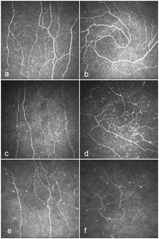
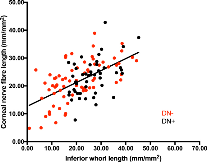
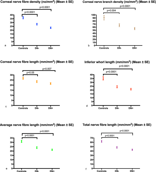
We assessed whether a measure of more distal corneal nerve fibre loss at the inferior whorl(IW) region is better than proximal measures of central corneal nerve damage in relation to the diagnosis of diabetic peripheral neuropathy(DPN), painful DPN and quality of life(QoL). Participants underwent detailed assessment of neuropathy, QoL using the SF36 questionnaire, pain visual analogue score(VAS), and corneal confocal microscopy(CCM). Corneal nerve fibre density (CNFD), branch density (CNBD) and length (CNFL) at the central cornea and inferior whorl length (IWL) and average(ANFL) and total(TNFL) nerve fibre length were compared in patients with and without DPN and between patients with and without painful DPN and in relation to QoL. All CCM parameters were significantly reduced, but IWL was reduced ~three-fold greater than CNFL in patients with and without DPN compared to controls. IWL(p = 0.001), ANFL(p = 0.01) and TNFL(p = 0.02) were significantly lower in patients with painful compared to painless DPN. The VAS score correlated with IWL(r = -0.36, P = 0.004), ANFL(r = -0.32, P = 0.01) and TNFL(r = -0.32, P = 0.01) and QoL correlated with CNFL(r = 0.35, P = 0.01) and IWL(r = 0.4, P = 0.004). Corneal nerve fibre damage is more prominent at the IW, lower in patients with painful compared to painless neuropathy and relates to their QoL. IWL may provide additional clinical utility for CCM in patients with DPN.

摘要

我们评估了在诊断糖尿病周围神经病变(DPN)、有痛性 DPN 和生活质量(QoL)方面,下象限(IW)区域的更远处角膜神经纤维损失测量值是否优于中央角膜神经损伤的近端测量值。参与者接受了详细的神经病变评估、使用 SF36 问卷评估 QoL、疼痛视觉模拟评分(VAS)以及角膜共聚焦显微镜(CCM)检查。比较了有和无 DPN 患者以及有和无有痛性 DPN 患者之间中央角膜和下象限长度(IWL)以及平均(ANFL)和总(TNFL)神经纤维长度的角膜神经纤维密度(CNFD)、分支密度(CNBD)和长度(CNFL)。所有 CCM 参数均显著降低,但与对照组相比,有和无 DPN 患者的 IWL 降低约三倍,而 CNFL 降低。与无痛性 DPN 相比,有痛性 DPN 患者的 IWL(p=0.001)、ANFL(p=0.01)和 TNFL(p=0.02)显著降低。VAS 评分与 IWL(r=-0.36,P=0.004)、ANFL(r=-0.32,P=0.01)和 TNFL(r=-0.32,P=0.01)呈负相关,QoL 与 CNFL(r=0.35,P=0.01)和 IWL(r=0.4,P=0.004)呈正相关。IW 处的角膜神经纤维损伤更为明显,有痛性 DPN 患者的神经纤维损伤程度较低,且与他们的 QoL 相关。IWL 可能为 DPN 患者的 CCM 提供额外的临床应用价值。

https://cdn.ncbi.nlm.nih.gov/pmc/blobs/5fb1/5818543/e6e6d03d4116/41598_2018_21643_Fig3_HTML.jpg
https://cdn.ncbi.nlm.nih.gov/pmc/blobs/5fb1/5818543/b2829144d76c/41598_2018_21643_Fig1_HTML.jpg
https://cdn.ncbi.nlm.nih.gov/pmc/blobs/5fb1/5818543/6daa8a2ced3d/41598_2018_21643_Fig2_HTML.jpg
https://cdn.ncbi.nlm.nih.gov/pmc/blobs/5fb1/5818543/e6e6d03d4116/41598_2018_21643_Fig3_HTML.jpg
https://cdn.ncbi.nlm.nih.gov/pmc/blobs/5fb1/5818543/b2829144d76c/41598_2018_21643_Fig1_HTML.jpg
https://cdn.ncbi.nlm.nih.gov/pmc/blobs/5fb1/5818543/6daa8a2ced3d/41598_2018_21643_Fig2_HTML.jpg
https://cdn.ncbi.nlm.nih.gov/pmc/blobs/5fb1/5818543/e6e6d03d4116/41598_2018_21643_Fig3_HTML.jpg

相似文献

1
Greater corneal nerve loss at the inferior whorl is related to the presence of diabetic neuropathy and painful diabetic neuropathy.下象限角膜神经损失越大与糖尿病神经病变和痛性糖尿病神经病变的存在有关。
Sci Rep. 2018 Feb 19;8(1):3283. doi: 10.1038/s41598-018-21643-z.
2
The Inferior Whorl For Detecting Diabetic Peripheral Neuropathy Using Corneal Confocal Microscopy.用于通过角膜共焦显微镜检测糖尿病周围神经病变的下旋轮。
Invest Ophthalmol Vis Sci. 2015 Apr;56(4):2498-504. doi: 10.1167/iovs.14-15919.
3
Corneal confocal microscopy for the diagnosis of diabetic peripheral neuropathy: A systematic review and meta-analysis.角膜共焦显微镜在糖尿病周围神经病变诊断中的应用:系统评价和荟萃分析。
J Diabetes Investig. 2022 Jan;13(1):134-147. doi: 10.1111/jdi.13643. Epub 2021 Aug 27.
4
Association of corneal nerve loss with markers of axonal ion channel dysfunction in type 1 diabetes.1 型糖尿病患者角膜神经丢失与轴突离子通道功能障碍标志物的关系。
Clin Neurophysiol. 2020 Jan;131(1):145-154. doi: 10.1016/j.clinph.2019.09.029. Epub 2019 Nov 11.
5
Corneal sub-basal whorl-like nerve plexus: a landmark for early and follow-up evaluation in transthyretin familial amyloid polyneuropathy.角膜基底层涡状神经丛:转甲状腺素蛋白家族性淀粉样多发性神经病早期和随访评估的标志。
Eur J Neurol. 2021 Feb;28(2):630-638. doi: 10.1111/ene.14563. Epub 2020 Oct 24.
6
Diabetic Neuropathy Is Characterized by Progressive Corneal Nerve Fiber Loss in the Central and Inferior Whorl Regions.糖尿病性神经病的特征是中央和下象限区域的角膜神经纤维渐进性丧失。
Invest Ophthalmol Vis Sci. 2020 Mar 9;61(3):48. doi: 10.1167/iovs.61.3.48.
7
Corneal confocal microscopy compared with quantitative sensory testing and nerve conduction for diagnosing and stratifying the severity of diabetic peripheral neuropathy.角膜共焦显微镜检查与定量感觉测试及神经传导检查在诊断和分层糖尿病性周围神经病变严重程度方面的比较
BMJ Open Diabetes Res Care. 2020 Dec;8(2). doi: 10.1136/bmjdrc-2020-001801.
8
Corneal confocal microscopy as a non-invasive test to assess diabetic peripheral neuropathy.角膜共焦显微镜作为一种非侵入性测试,用于评估糖尿病周围神经病变。
Diabetes Res Clin Pract. 2018 Feb;136:85-92. doi: 10.1016/j.diabres.2017.11.026. Epub 2017 Dec 6.
9
Small fiber neuropathy for assessment of disease severity in amyotrophic lateral sclerosis: corneal confocal microscopy findings.用于评估肌萎缩侧索硬化症疾病严重程度的小纤维神经病:角膜共聚焦显微镜检查结果。
Orphanet J Rare Dis. 2022 Jan 6;17(1):7. doi: 10.1186/s13023-021-02157-w.
10
Corneal confocal microscopy detects small nerve fibre damage in patients with painful diabetic neuropathy.角膜共焦显微镜检测痛性糖尿病周围神经病患者的小纤维神经损伤。
Sci Rep. 2020 Feb 25;10(1):3371. doi: 10.1038/s41598-020-60422-7.

引用本文的文献

1
Mechanisms of Corneal Nerve Regeneration: Examining Molecular Regulators.角膜神经再生机制:探究分子调节因子
Cells. 2025 Aug 27;14(17):1322. doi: 10.3390/cells14171322.
2
Machine Learning to Diagnose Complications of Diabetes.用于诊断糖尿病并发症的机器学习
J Diabetes Sci Technol. 2025 Sep 11:19322968251365245. doi: 10.1177/19322968251365245.
3
Methods for the evaluation of corneal nerve fibres in diabetes mellitus by in vivo confocal microscopy: a scoping review.通过体内共聚焦显微镜评估糖尿病患者角膜神经纤维的方法:一项范围综述

本文引用的文献

1
Corneal confocal microscopy is a rapid reproducible ophthalmic technique for quantifying corneal nerve abnormalities.角膜共焦显微镜检查是一种用于量化角膜神经异常的快速可重复的眼科技术。
PLoS One. 2017 Aug 17;12(8):e0183040. doi: 10.1371/journal.pone.0183040. eCollection 2017.
2
Diagnostic utility of corneal confocal microscopy and intra-epidermal nerve fibre density in diabetic neuropathy.角膜共焦显微镜检查和表皮内神经纤维密度在糖尿病神经病变中的诊断效用
PLoS One. 2017 Jul 18;12(7):e0180175. doi: 10.1371/journal.pone.0180175. eCollection 2017.
3
Use of Corneal Confocal Microscopy to Evaluate Small Nerve Fibers in Patients With Human Immunodeficiency Virus.
BMJ Open. 2025 Sep 4;15(9):e098294. doi: 10.1136/bmjopen-2024-098294.
4
Corneal Nerve Morphology in Painful Diabetic Neuropathy: A Meta-Analysis of In Vivo Confocal Microscopy Studies.疼痛性糖尿病神经病变中的角膜神经形态学:一项共聚焦显微镜活体研究的荟萃分析
Biomedicines. 2025 Jul 8;13(7):1675. doi: 10.3390/biomedicines13071675.
5
Cornea Nerves Can Identify Different Types of Parkinson's Disease.角膜神经可识别不同类型的帕金森病。
Invest Ophthalmol Vis Sci. 2025 Jun 2;66(6):21. doi: 10.1167/iovs.66.6.21.
6
Automated program using convolutional neural networks for objective and reproducible selection of corneal confocal microscopy images.使用卷积神经网络的自动化程序用于角膜共焦显微镜图像的客观且可重复选择。
Digit Health. 2025 Mar 17;11:20552076251326223. doi: 10.1177/20552076251326223. eCollection 2025 Jan-Dec.
7
Geometrical Features of Subbasal Corneal Whorl-like Nerve Patterns in Dry Eye Disease: An In Vivo Confocal Microscopy Study.干眼疾病中角膜基底膜下漩涡状神经模式的几何特征:一项活体共聚焦显微镜研究
Ophthalmol Sci. 2024 Dec 5;5(2):100669. doi: 10.1016/j.xops.2024.100669. eCollection 2025 Mar-Apr.
8
Small fiber neuropathy in the post-COVID condition and Myalgic Encephalomyelitis/Chronic Fatigue Syndrome: Clinical significance and diagnostic challenges.新冠后状态下的小纤维神经病变与肌痛性脑脊髓炎/慢性疲劳综合征:临床意义及诊断挑战
Eur J Neurol. 2025 Feb;32(2):e70016. doi: 10.1111/ene.70016.
9
Deep learning-based automated tool for diagnosing diabetic peripheral neuropathy.基于深度学习的糖尿病周围神经病变诊断自动化工具。
Digit Health. 2024 Dec 25;10:20552076241307573. doi: 10.1177/20552076241307573. eCollection 2024 Jan-Dec.
10
Quantifying the Corneal Nerve Whorl Pattern.量化角膜神经螺旋模式。
Transl Vis Sci Technol. 2024 Dec 2;13(12):11. doi: 10.1167/tvst.13.12.11.
使用角膜共焦显微镜评估人类免疫缺陷病毒患者的小神经纤维。
JAMA Ophthalmol. 2017 Jul 1;135(7):795-800. doi: 10.1001/jamaophthalmol.2017.1703.
4
Effect of omega-3 supplementation on neuropathy in type 1 diabetes: A 12-month pilot trial.ω-3补充剂对1型糖尿病神经病变的影响:一项为期12个月的试点试验。
Neurology. 2017 Jun 13;88(24):2294-2301. doi: 10.1212/WNL.0000000000004033. Epub 2017 May 17.
5
Cibinetide Improves Corneal Nerve Fiber Abundance in Patients With Sarcoidosis-Associated Small Nerve Fiber Loss and Neuropathic Pain.西贝肽可改善结节病相关小神经纤维损伤和神经性疼痛患者的角膜神经纤维丰度。
Invest Ophthalmol Vis Sci. 2017 May 1;58(6):BIO52-BIO60. doi: 10.1167/iovs.16-21291.
6
The Pain in Neuropathy Study (PiNS): a cross-sectional observational study determining the somatosensory phenotype of painful and painless diabetic neuropathy.神经病变疼痛研究(PiNS):一项横断面观察性研究,旨在确定疼痛性和无痛性糖尿病神经病变的躯体感觉表型。
Pain. 2016 May;157(5):1132-1145. doi: 10.1097/j.pain.0000000000000491.
7
Corneal confocal microscopy in chronic inflammatory demyelinating polyneuropathy.慢性炎症性脱髓鞘性多发性神经病的角膜共焦显微镜检查。
Ann Clin Transl Neurol. 2015 Dec 28;3(2):88-100. doi: 10.1002/acn3.275. eCollection 2016 Feb.
8
Corneal Confocal Microscopy Detects Small Fibre Neuropathy in Patients with Upper Gastrointestinal Cancer and Nerve Regeneration in Chemotherapy Induced Peripheral Neuropathy.角膜共聚焦显微镜检测上消化道癌患者的小纤维神经病变及化疗所致周围神经病变中的神经再生
PLoS One. 2015 Oct 2;10(10):e0139394. doi: 10.1371/journal.pone.0139394. eCollection 2015.
9
Imaging of the Corneal Subbasal Whorl-like Nerve Plexus: More Accurate Depiction of the Extent of Corneal Nerve Damage in Patients With Diabetes.角膜基底膜下涡状神经丛成像:更准确描绘糖尿病患者角膜神经损伤程度
Invest Ophthalmol Vis Sci. 2015 Aug;56(9):5417-23. doi: 10.1167/iovs.15-16609.
10
Utility of Assessing Nerve Morphology in Central Cornea Versus Whorl Area for Diagnosing Diabetic Peripheral Neuropathy.评估中央角膜与涡状区域神经形态在诊断糖尿病周围神经病变中的效用。
Cornea. 2015 Jul;34(7):756-61. doi: 10.1097/ICO.0000000000000447.