Li Siwei, Tallia Francesca, Mohammed Ali A, Stevens Molly M, Jones Julian R
Department of Materials, Imperial College London, South Kensington Campus, London, SW7 2AZ, UK.
Biomater Sci. 2020 Aug 21;8(16):4458-4466. doi: 10.1039/c9bm01829h. Epub 2020 Feb 26.
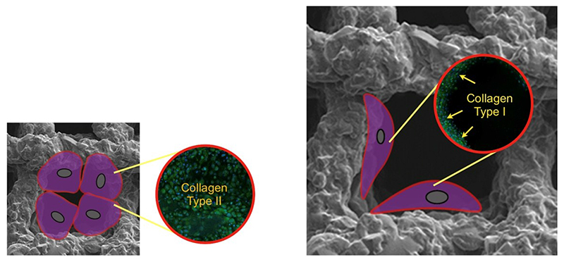
We report that 3-D printed scaffold channel size can direct bone marrow derived stem cell differentiation. Treatment of articular cartilage trauma injuries, such as microfracture surgery, have limited success because durability is limited as fibrocartilage forms. A scaffold-assisted approach, combining microfracture with biomaterials has potential if the scaffold can promote articular cartilage production and share load with cartilage. Here, we investigated human bone marrow derived stromal cell (hBMSC) differentiation in vitro in 3-D printed silica/poly(tetrahydrofuran)/poly(ε-caprolactone) hybrid scaffolds with specific channel sizes. Channel widths of ∼230 μm (210 ± 22 μm mean strut size, 42.4 ± 3.9% porosity) provoked hBMSC differentiation down a chondrogenic path, with collagen Type II matrix prevalent, indicative of hyaline cartilage. When pores were larger (∼500 μm, 229 ± 29 μm mean strut size, 63.8 ± 1.6% porosity) collagen Type I was dominant, indicating fibrocartilage. There was less matrix and voids in smaller channels (∼100 μm, 218 ± 28 μm mean strut size, 31.2 ± 2.9% porosity). Our findings suggest that a 200-250 μm pore channel width, in combination with the surface chemistry and stiffness of the scaffold, is optimal for cell-cell interactions to promote chondrogenic differentiation and enable the chondrocytes to maintain their phenotype.
我们报告称,3D打印支架的通道大小可引导骨髓来源干细胞的分化。治疗关节软骨创伤性损伤,如微骨折手术,成功率有限,因为随着纤维软骨形成,其耐久性受到限制。如果支架能够促进关节软骨生成并与软骨分担负荷,那么将微骨折与生物材料相结合的支架辅助方法具有潜力。在此,我们研究了人骨髓来源基质细胞(hBMSC)在具有特定通道大小的3D打印二氧化硅/聚(四氢呋喃)/聚(ε-己内酯)混合支架中的体外分化情况。约230μm的通道宽度(平均支柱尺寸210±22μm,孔隙率42.4±3.9%)促使hBMSC向软骨生成方向分化,II型胶原基质普遍存在,表明为透明软骨。当孔隙较大时(约500μm,平均支柱尺寸229±29μm,孔隙率63.8±1.6%),I型胶原占主导,表明为纤维软骨。较小通道(约100μm,平均支柱尺寸218±28μm,孔隙率31.2±2.9%)中的基质和空隙较少。我们的研究结果表明,200 - 250μm的孔隙通道宽度,结合支架的表面化学性质和硬度,最有利于细胞间相互作用,促进软骨生成分化,并使软骨细胞维持其表型。