Fourth Department of Internal Medicine, Teikyo University Mizonokuchi Hospital, 5-1-1 Futako, Takatsu-ku, Kawasaki-shi, Kanagawa, 213-8507, Japan.
Department of Pathology and Laboratory Medicine, Institute of Biomedical Sciences, Tokushima University Graduate School, Tokushima-shi, Tokushima, Japan.
BMC Gastroenterol. 2020 Feb 27;20(1):46. doi: 10.1186/s12876-020-01194-2.
Non-alcoholic fatty liver disease (NAFLD) is a hepatic manifestation of metabolic syndrome. Within the spectrum of NAFLD, non-alcoholic steatohepatitis (NASH) in combination with hepatic inflammation and fibrosis can lead to liver cirrhosis and hepatocellular carcinoma. Dysbiosis was reported to contribute to NASH pathogenesis. This study aimed to determine the effects of fructo-oligosaccharides (FOS) on steatohepatitis and visceral adiposity in an obese mouse model of NASH.
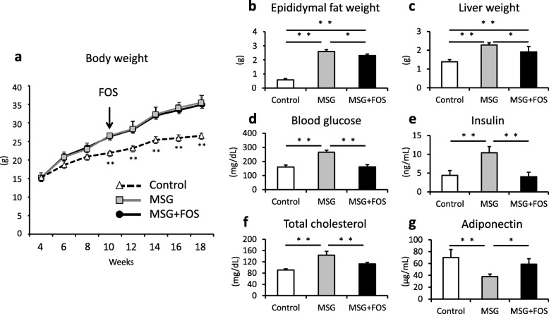
Twelve newborn C57BL/6 J male mice were subcutaneously injected with monosodium glutamate (MSG) to induce obesity on a conventional diet. Six mice were also administered 5% FOS via drinking water from 10 weeks of age. At 18 weeks, histological characteristics of the liver and epididymal fat were compared between the groups. Hepatic mRNA expression of lipid metabolism enzymes and SCFA in feces and sera were measured.
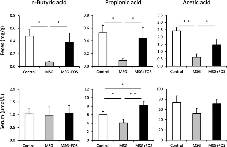
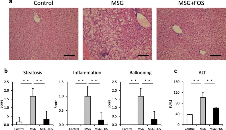
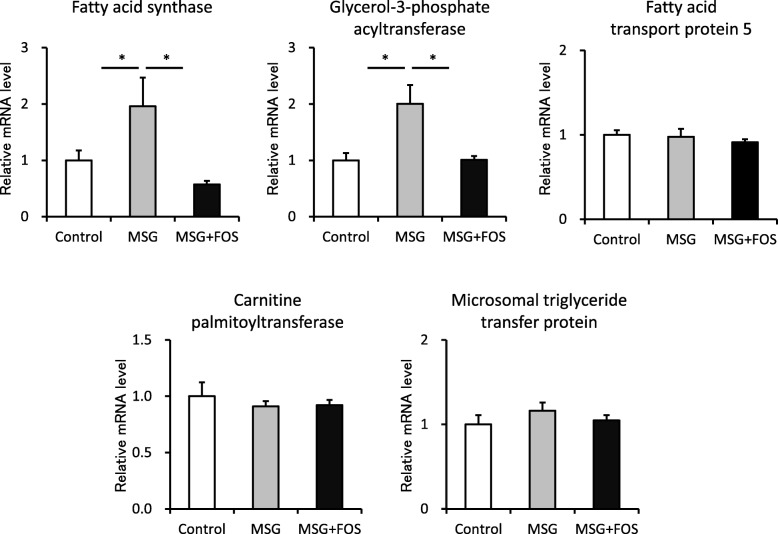
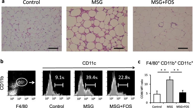
Hepatic steatosis, inflammatory cell infiltration, and hepatocyte ballooning in the liver and increased hepatic mRNA expression of fatty acid synthase and glycerol-3-phosphate acyltransferase were observed in the MSG-treated mice. FOS treatment improved the liver pathology and blunted the increases in the mRNA expression levels of lipid metabolism enzymes. In addition, FOS inhibited adipocyte enlargement and formation of crown-like structures and reduced the M1 macrophage frequency in the epididymal fat of the MSG mice (39.4% ± 3.0% vs. 22.8% ± 0.7%; P = 0.001). FOS increased not only the fecal concentrations of n-butyric acid (0.04 ± 0.01 vs. 0.38 ± 0.14 mg/g, P = 0.02), propionic acid (0.09 ± 0.03 vs. 0.42 ± 0.16 mg/g, P = 0.02), and acetic acid (0.65 ± 0.16 vs. 1.48 ± 0.29 mg/g, P = 0.03) but also the serum concentration of propionic acid (3.9 ± 0.5 vs. 8.2 ± 0.5 μmol/L, P = 0.001).
FOS ameliorates steatohepatitis, visceral adiposity, and chronic inflammation by increasing SCFA production.
非酒精性脂肪性肝病(NAFLD)是代谢综合征的肝脏表现。在 NAFLD 谱中,非酒精性脂肪性肝炎(NASH)合并肝炎症和纤维化可导致肝硬化和肝细胞癌。肠道菌群失调被报道与 NASH 的发病机制有关。本研究旨在确定果寡糖(FOS)对 NASH 肥胖小鼠模型中脂肪性肝炎和内脏脂肪堆积的影响。
将 12 只新生 C57BL/6J 雄性小鼠皮下注射谷氨酸单钠以诱导肥胖,同时给予常规饮食。从 10 周龄开始,6 只小鼠还通过饮用水给予 5% FOS。在 18 周时,比较各组肝脏和附睾脂肪的组织学特征。测量粪便和血清中脂质代谢酶和短链脂肪酸(SCFA)的肝 mRNA 表达。
在 MSG 处理的小鼠中观察到肝脏脂肪变性、炎症细胞浸润和肝细胞气球样变,以及肝脂肪酸合成酶和甘油-3-磷酸酰基转移酶 mRNA 表达增加。FOS 治疗改善了肝病理,并抑制了脂质代谢酶 mRNA 表达水平的升高。此外,FOS 抑制了脂肪细胞增大和冠层结构的形成,并减少了 MSG 小鼠附睾脂肪中 M1 巨噬细胞的频率(39.4%±3.0%比 22.8%±0.7%;P=0.001)。FOS 不仅增加了粪便中丁酸(0.04±0.01 比 0.38±0.14mg/g,P=0.02)、丙酸(0.09±0.03 比 0.42±0.16mg/g,P=0.02)和乙酸(0.65±0.16 比 1.48±0.29mg/g,P=0.03)的浓度,还增加了血清中丙酸的浓度(3.9±0.5 比 8.2±0.5μmol/L,P=0.001)。
FOS 通过增加 SCFA 的产生来改善脂肪性肝炎、内脏脂肪堆积和慢性炎症。