The Henry Royce Institute and School of Materials, The University of Manchester, Manchester, M13 9PL, UK.
BMC Biol. 2020 Feb 27;18(1):21. doi: 10.1186/s12915-020-0753-2.
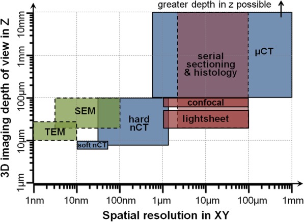
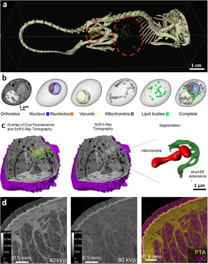
Recent developments within micro-computed tomography (μCT) imaging have combined to extend our capacity to image tissue in three (3D) and four (4D) dimensions at micron and sub-micron spatial resolutions, opening the way for virtual histology, live cell imaging, subcellular imaging and correlative microscopy. Pivotal to this has been the development of methods to extend the contrast achievable for soft tissue. Herein, we review the new capabilities within the field of life sciences imaging, and consider how future developments in this field could further benefit the life sciences community.
最近在微计算机断层扫描(μCT)成像方面的发展结合在一起,扩展了我们在微米和亚微米空间分辨率下以三维(3D)和四维(4D)成像组织的能力,为虚拟组织学、活细胞成像、亚细胞成像和相关显微镜成像开辟了道路。关键是开发了扩展软组织可实现对比度的方法。本文综述了生命科学成像领域的新功能,并探讨了该领域未来的发展如何使生命科学领域受益。