Bleyer Martina, Kunze Marius, Gruber-Dujardin Eva, Mätz-Rensing Kerstin
Pathology Unit, German Primate Center, Kellnerweg 4, 37077 Göttingen, Germany.
Primate Biol. 2017 Mar 1;4(1):17-25. doi: 10.5194/pb-4-17-2017. eCollection 2017.
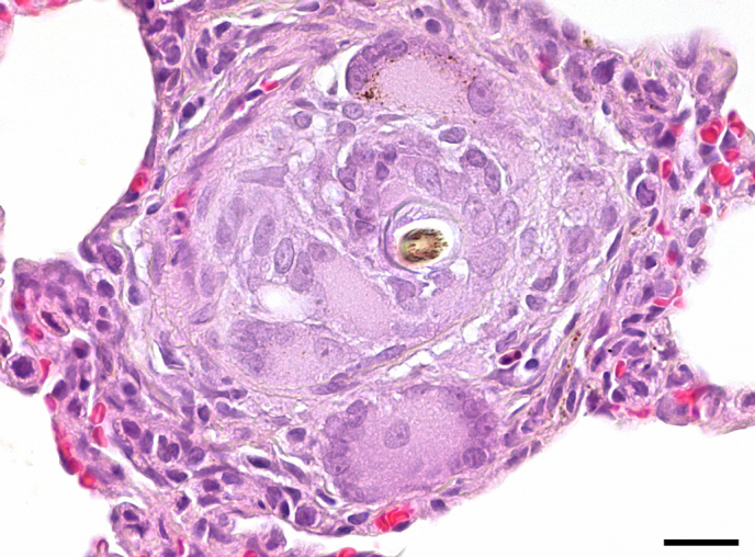
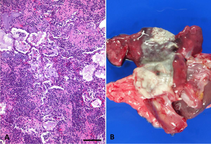
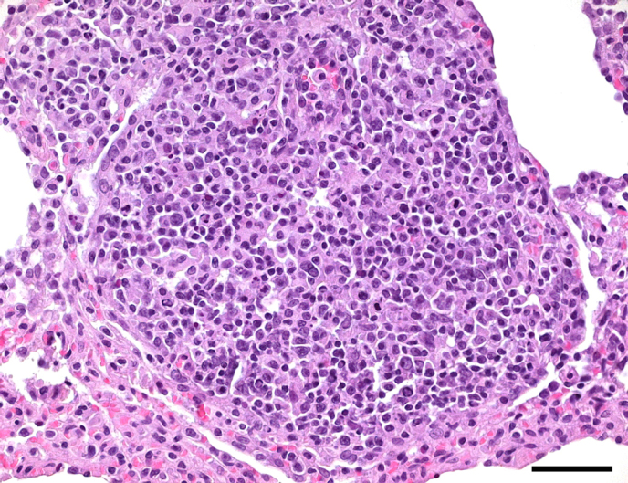
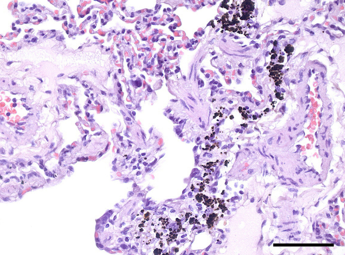
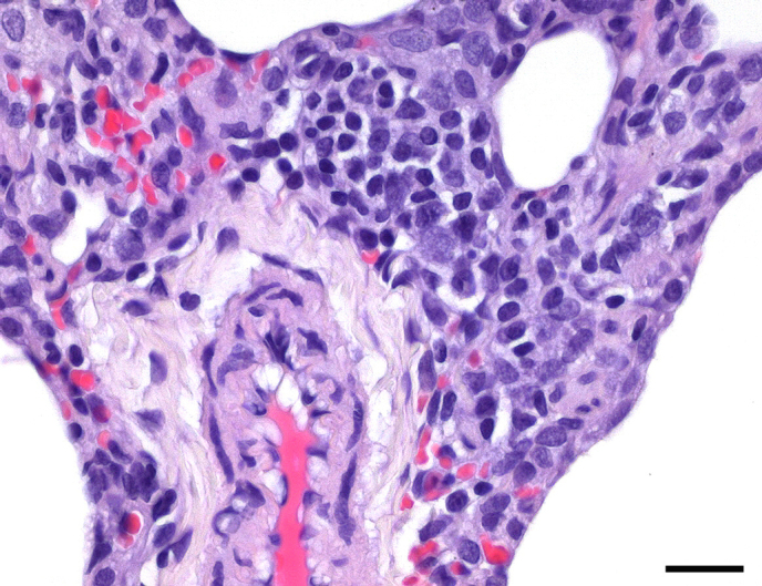
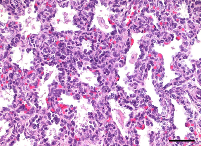
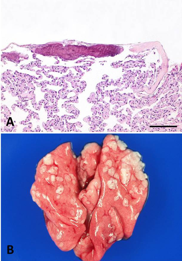
Data on spontaneous pathology are substantially scarce for common marmosets, compared to other laboratory animals, but is essential for the interpretation of histological findings in the context of toxicological and experimental studies. Especially if common marmosets are used as experimental animals in respiratory research, detailed knowledge on the spectrum, occurrence, and incidence of spontaneous histopathological pulmonary lesions in this non-human primate species is required. In this study, lung tissue of 638 common marmosets from the marmoset colony of the German Primate Center was examined histologically. The analysis revealed a high incidence of predominantly mild and multifocal interstitial pneumonia (32.99 %) of unknown etiology in most cases. Only few marmosets exhibited lobar pneumonia (1.41 %) and bronchopneumonia (0.94), which were mainly caused by bacterial pathogens such as and . Lung immaturity and atelectasis were common histological findings in newborn marmosets. Typical background lesions included anthracosis (8.15 %), hemosiderosis (1.72 %), extramedullary hematopoiesis (11.6 %), mineralization (10.97 %), and inflammatory cell foci (10.34 %). In addition, three cases of pulmonary arteriopathy (0.47 %) and 1 case of foreign-body granuloma (0.16 %) were detected in the marmoset study cohort. The high prevalence of circulatory disturbances (congestion, edema, hemorrhage) and changes in air content (secondary atelectasis, alveolar emphysema) could partly be explained by euthanasia-related artifacts or agonal changes. The present study provides a comprehensive overview of the range and incidence of spontaneous pulmonary histopathology in common marmosets, serving as valuable reference data for the interpretation of lung lesions in toxicological and experimental marmoset studies.
与其他实验动物相比,普通狨猴自发性病理的数据非常稀少,但对于在毒理学和实验研究背景下解释组织学结果至关重要。特别是当普通狨猴被用作呼吸研究的实验动物时,需要详细了解这种非人灵长类动物自发性组织病理学肺部病变的范围、发生情况和发病率。在本研究中,对来自德国灵长类动物中心狨猴种群的638只普通狨猴的肺组织进行了组织学检查。分析显示,在大多数情况下,主要为轻度和多灶性间质性肺炎的发病率很高(32.99%),病因不明。只有少数狨猴表现出大叶性肺炎(1.41%)和支气管肺炎(0.94%),主要由 和 等细菌病原体引起。肺不成熟和肺不张是新生狨猴常见的组织学表现。典型的背景病变包括煤尘肺(8.15%)、含铁血黄素沉着症(1.72%)、髓外造血(11.6%)、矿化(10.97%)和炎症细胞灶(10.34%)。此外,在狨猴研究队列中检测到3例肺动脉病(0.47%)和1例异物肉芽肿(0.16%)。循环系统紊乱(充血、水肿、出血)和空气含量变化(继发性肺不张、肺泡气肿)的高发生率部分可以由安乐死相关的伪像或濒死变化来解释。本研究全面概述了普通狨猴自发性肺部组织病理学的范围和发病率,为解释毒理学和实验性狨猴研究中的肺部病变提供了有价值的参考数据。