Payabvash Seyedmehdi, Aboian Mariam, Tihan Tarik, Cha Soonmee
Department of Radiology and Biomedical Imaging, Yale School of Medicine, New Haven, CT, United States.
Department of Radiology and Biomedical Imaging, University of California, San Francisco, San Francisco, CA, United States.
Front Oncol. 2020 Feb 7;10:71. doi: 10.3389/fonc.2020.00071. eCollection 2020.
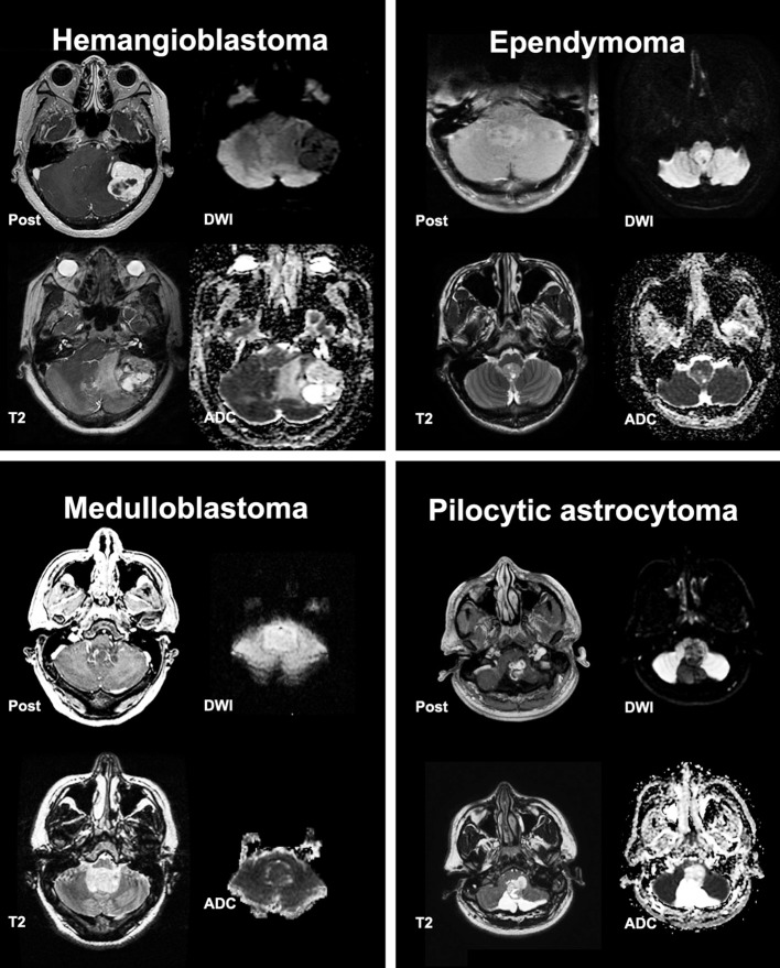
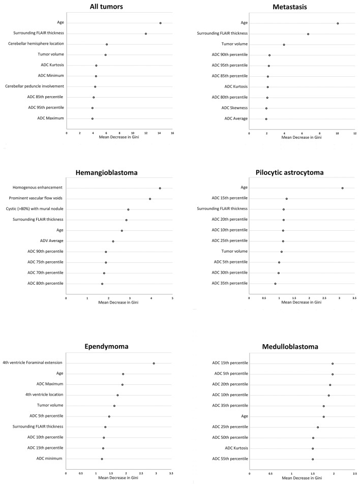
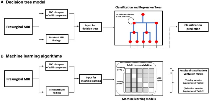
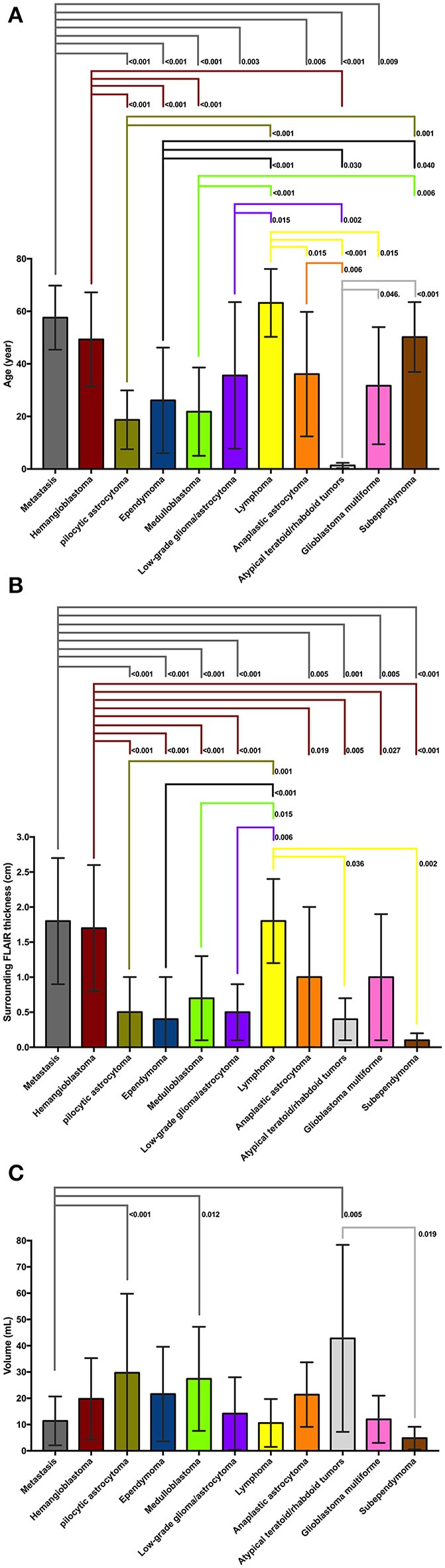
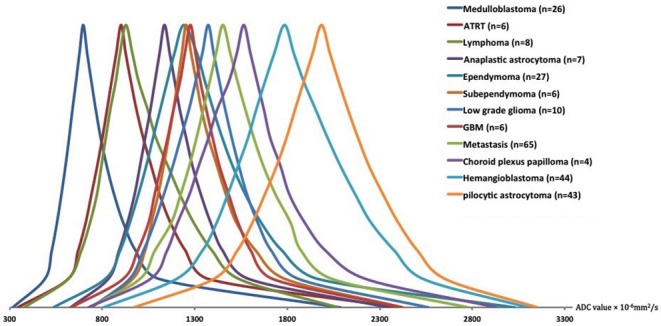
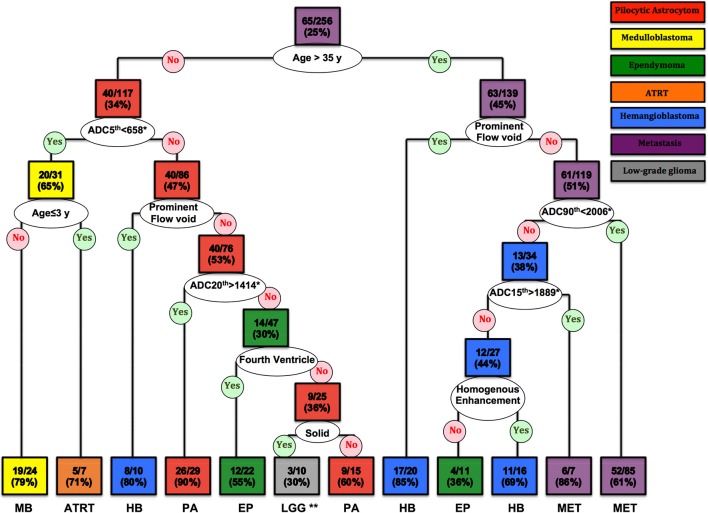
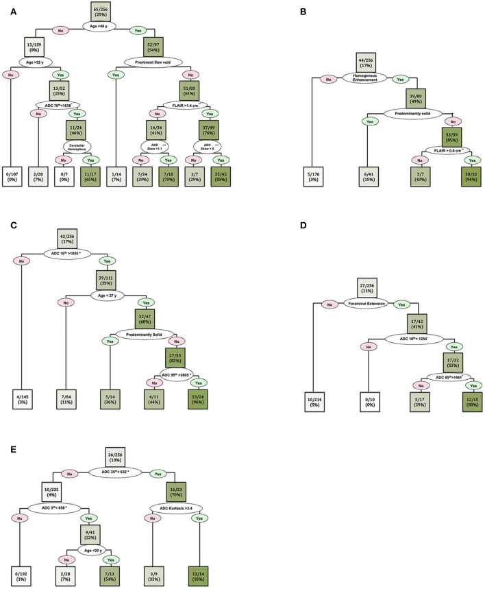
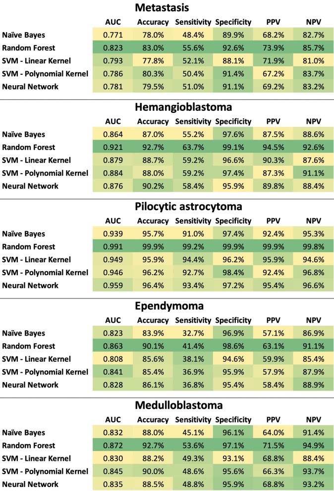
We applied machine learning algorithms for differentiation of posterior fossa tumors using apparent diffusion coefficient (ADC) histogram analysis and structural MRI findings. A total of 256 patients with intra-axial posterior fossa tumors were identified, of whom 248 were included in machine learning analysis, with at least 6 representative subjects per each tumor pathology. The ADC histograms of solid components of tumors, structural MRI findings, and patients' age were applied to construct decision models using Classification and Regression Tree analysis. We also compared different machine learning classification algorithms (i.e., naïve Bayes, random forest, neural networks, support vector machine with linear and polynomial kernel) for dichotomized differentiation of the 5 most common tumors in our cohort: metastasis ( = 65), hemangioblastoma ( = 44), pilocytic astrocytoma ( = 43), ependymoma ( = 27), and medulloblastoma ( = 26). The decision tree model could differentiate seven tumor histopathologies with terminal nodes yielding up to 90% accurate classification rates. In receiver operating characteristics (ROC) analysis, the decision tree model achieved greater area under the curve (AUC) for differentiation of pilocytic astrocytoma ( = 0.020); and atypical teratoid/rhabdoid tumor ATRT ( = 0.001) from other types of neoplasms compared to the official clinical report. However, neuroradiologists' interpretations had greater accuracy in differentiating metastases ( = 0.001). Among different machine learning algorithms, random forest models yielded the highest accuracy in dichotomized classification of the 5 most common tumor types; and in multiclass differentiation of all tumor types random forest yielded an averaged AUC of 0.961 in training datasets, and 0.873 in validation samples. Our study demonstrates the potential application of machine learning algorithms and decision trees for accurate differentiation of brain tumors based on pretreatment MRI. Using easy to apply and understandable imaging metrics, the proposed decision tree model can help radiologists with differentiation of posterior fossa tumors, especially in tumors with similar qualitative imaging characteristics. In particular, our decision tree model provided more accurate differentiation of pilocytic astrocytomas from ATRT than by neuroradiologists in clinical reads.
我们应用机器学习算法,通过表观扩散系数(ADC)直方图分析和结构磁共振成像(MRI)结果来鉴别后颅窝肿瘤。共识别出256例轴内后颅窝肿瘤患者,其中248例纳入机器学习分析,每种肿瘤病理类型至少有6名代表性受试者。将肿瘤实性成分的ADC直方图、结构MRI结果和患者年龄应用于使用分类与回归树分析构建决策模型。我们还比较了不同的机器学习分类算法(即朴素贝叶斯、随机森林、神经网络、具有线性和多项式核的支持向量机),用于对我们队列中5种最常见肿瘤进行二分法鉴别:转移瘤(n = 65)、血管母细胞瘤(n = 44)、毛细胞型星形细胞瘤(n = 43)、室管膜瘤(n = 27)和髓母细胞瘤(n = 26)。决策树模型能够鉴别7种肿瘤组织病理学类型,其终端节点的分类准确率高达90%。在受试者操作特征(ROC)分析中,与官方临床报告相比,决策树模型在鉴别毛细胞型星形细胞瘤(AUC = 0.920)和非典型畸胎样/横纹肌样肿瘤(ATRT,AUC = 0.901)与其他类型肿瘤方面,曲线下面积(AUC)更大。然而,神经放射科医生的解读在鉴别转移瘤方面准确性更高(AUC = 0.901)。在不同的机器学习算法中,随机森林模型在对5种最常见肿瘤类型进行二分法分类时准确率最高;在对所有肿瘤类型进行多分类鉴别时,随机森林在训练数据集中的平均AUC为0.961,在验证样本中的平均AUC为0.873。我们的研究证明了机器学习算法和决策树在基于治疗前MRI准确鉴别脑肿瘤方面的潜在应用。使用易于应用和理解的成像指标,所提出的决策树模型可以帮助放射科医生鉴别后颅窝肿瘤,特别是在具有相似定性成像特征的肿瘤中。特别是,我们的决策树模型在临床读片中比神经放射科医生更准确地鉴别了毛细胞型星形细胞瘤和ATRT。