• 文献检索
  • 文档翻译
  • 深度研究
  • 学术资讯
  • Suppr Zotero 插件Zotero 插件
  • 邀请有礼
  • 套餐&价格
  • 历史记录
应用&插件
Suppr Zotero 插件Zotero 插件浏览器插件Mac 客户端Windows 客户端微信小程序
定价
高级版会员购买积分包购买API积分包
服务
文献检索文档翻译深度研究API 文档MCP 服务
关于我们
关于 Suppr公司介绍联系我们用户协议隐私条款
关注我们

Suppr 超能文献

核心技术专利:CN118964589B侵权必究
粤ICP备2023148730 号-1Suppr @ 2026

文献检索

告别复杂PubMed语法,用中文像聊天一样搜索,搜遍4000万医学文献。AI智能推荐,让科研检索更轻松。

立即免费搜索

文件翻译

保留排版,准确专业,支持PDF/Word/PPT等文件格式,支持 12+语言互译。

免费翻译文档

深度研究

AI帮你快速写综述,25分钟生成高质量综述,智能提取关键信息,辅助科研写作。

立即免费体验

肌软:一种使用机器学习算法的自动化肌肉组织学分析工具,利用 FIJI/ImageJ 软件。

Myosoft: An automated muscle histology analysis tool using machine learning algorithm utilizing FIJI/ImageJ software.

机构信息

Department of Cell Biology, School of Medicine, Emory University, Atlanta, Georgia, United States of America.

Undergraduate program in Neuroscience and Behavioral Biology, School of Medicine, Emory University, Atlanta, Georgia, United States of America.

出版信息

PLoS One. 2020 Mar 4;15(3):e0229041. doi: 10.1371/journal.pone.0229041. eCollection 2020.

DOI:10.1371/journal.pone.0229041
PMID:32130242
原文链接:https://pmc.ncbi.nlm.nih.gov/articles/PMC7055860/
Abstract

METHODS

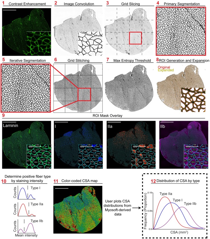
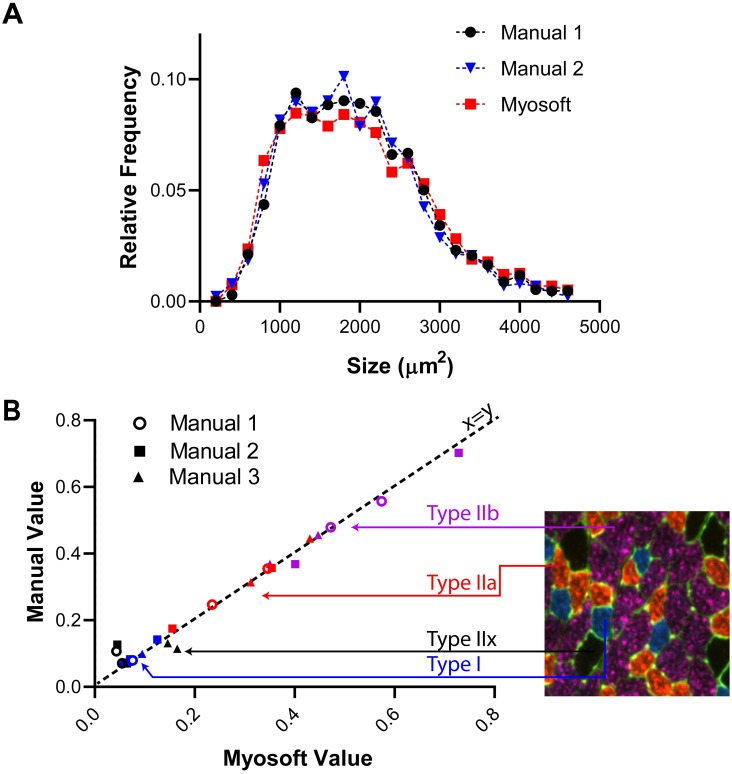
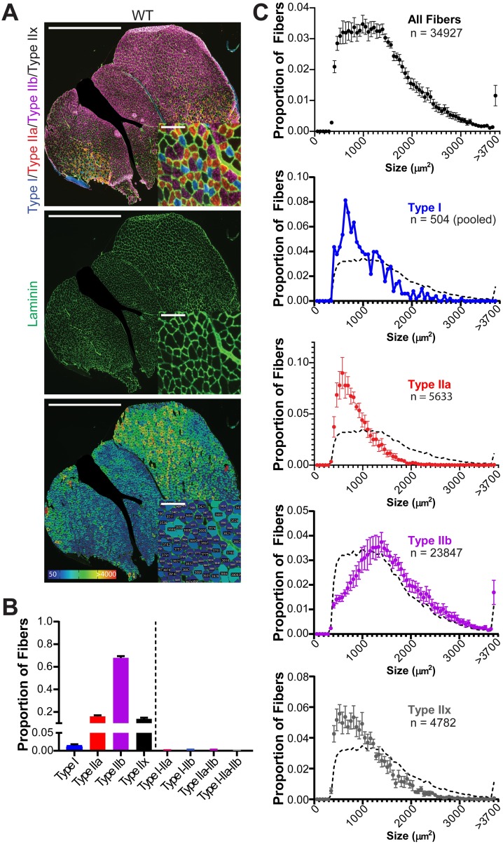
Muscle sections were stained for cell boundary (laminin) and myofiber type (myosin heavy chain isoforms). Myosoft, running in the open access software platform FIJI (ImageJ), was used to analyze myofiber size and type in transverse sections of entire gastrocnemius/soleus muscles.

RESULTS

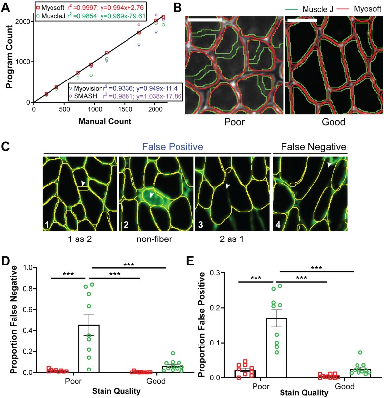
Myosoft provides an accurate analysis of hundreds to thousands of muscle fibers within 25 minutes, which is >10-times faster than manual analysis. We demonstrate that Myosoft is capable of handling high-content images even when image or staining quality is suboptimal, which is a marked improvement over currently available and comparable programs.

CONCLUSIONS

Myosoft is a reliable, accurate, high-throughput, and convenient tool to analyze high-content muscle histology. Myosoft is freely available to download from Github at https://github.com/Hyojung-Choo/Myosoft/tree/Myosoft-hub.

摘要

方法

肌肉切片经细胞边界(层粘连蛋白)和肌纤维类型(肌球蛋白重链同工型)染色。Myosoft 运行在开放获取的软件平台 FIJI(ImageJ)上,用于分析整个比目鱼肌/跟腱肌肉横切面上的肌纤维大小和类型。

结果

Myosoft 能够在 25 分钟内对数百到数千条肌纤维进行精确分析,比手动分析快 10 多倍。我们证明,即使在图像或染色质量不理想的情况下,Myosoft 也能够处理高内涵图像,这比当前可用的和可比的程序有显著的改进。

结论

Myosoft 是一种可靠、准确、高通量和方便的分析高内涵肌肉组织学的工具。Myosoft 可从 Github 上免费下载,网址为 https://github.com/Hyojung-Choo/Myosoft/tree/Myosoft-hub。

https://cdn.ncbi.nlm.nih.gov/pmc/blobs/2dc2/7055860/c151af12cda7/pone.0229041.g007.jpg
https://cdn.ncbi.nlm.nih.gov/pmc/blobs/2dc2/7055860/39a835665462/pone.0229041.g001.jpg
https://cdn.ncbi.nlm.nih.gov/pmc/blobs/2dc2/7055860/7d580d794761/pone.0229041.g002.jpg
https://cdn.ncbi.nlm.nih.gov/pmc/blobs/2dc2/7055860/f16b711e59e6/pone.0229041.g003.jpg
https://cdn.ncbi.nlm.nih.gov/pmc/blobs/2dc2/7055860/178e865e1e8f/pone.0229041.g004.jpg
https://cdn.ncbi.nlm.nih.gov/pmc/blobs/2dc2/7055860/bedf6733051e/pone.0229041.g005.jpg
https://cdn.ncbi.nlm.nih.gov/pmc/blobs/2dc2/7055860/0342522b02fd/pone.0229041.g006.jpg
https://cdn.ncbi.nlm.nih.gov/pmc/blobs/2dc2/7055860/c151af12cda7/pone.0229041.g007.jpg

相似文献

1
Myosoft: An automated muscle histology analysis tool using machine learning algorithm utilizing FIJI/ImageJ software.肌软:一种使用机器学习算法的自动化肌肉组织学分析工具,利用 FIJI/ImageJ 软件。
PLoS One. 2020 Mar 4;15(3):e0229041. doi: 10.1371/journal.pone.0229041. eCollection 2020.
2
MyoSight-semi-automated image analysis of skeletal muscle cross sections.MyoSight—骨骼肌横切片的半自动图像分析。
Skelet Muscle. 2020 Nov 16;10(1):33. doi: 10.1186/s13395-020-00250-5.
3
Open-CSAM, a new tool for semi-automated analysis of myofiber cross-sectional area in regenerating adult skeletal muscle.Open-CSAM,一种用于分析再生成年骨骼肌肌纤维横截面积的半自动新工具。
Skelet Muscle. 2019 Jan 8;9(1):2. doi: 10.1186/s13395-018-0186-6.
4
MuscleJ: a high-content analysis method to study skeletal muscle with a new Fiji tool.MuscleJ:一种使用新 Fiji 工具研究骨骼肌的高内涵分析方法。
Skelet Muscle. 2018 Aug 6;8(1):25. doi: 10.1186/s13395-018-0171-0.
5
Automatic and unbiased segmentation and quantification of myofibers in skeletal muscle.骨骼肌中原纤维的自动和无偏分割与定量。
Sci Rep. 2021 Jun 3;11(1):11793. doi: 10.1038/s41598-021-91191-6.
6
Automated muscle histopathology analysis using CellProfiler.使用 CellProfiler 进行自动化肌肉组织病理学分析。
Skelet Muscle. 2018 Oct 18;8(1):32. doi: 10.1186/s13395-018-0178-6.
7
Automated image segmentation method to analyse skeletal muscle cross section in exercise-induced regenerating myofibers.自动化图像分割方法分析运动诱导再生肌纤维的骨骼肌横截面积。
Sci Rep. 2021 Oct 29;11(1):21327. doi: 10.1038/s41598-021-00886-3.
8
INSIDIA: A FIJI Macro Delivering High-Throughput and High-Content Spheroid Invasion Analysis.INSIDIA:一种斐济宏病毒,可实现高通量和高内涵球体侵袭分析。
Biotechnol J. 2017 Oct;12(10). doi: 10.1002/biot.201700140. Epub 2017 Sep 6.
9
Automated image analysis of skeletal muscle fiber cross-sectional area.骨骼肌纤维横截面积的自动化图像分析。
J Appl Physiol (1985). 2013 Jan 1;114(1):148-55. doi: 10.1152/japplphysiol.01022.2012. Epub 2012 Nov 8.
10
Approach for semi-automated measurement of fiber diameter in murine and canine skeletal muscle.用于在鼠和犬骨骼肌中半自动测量纤维直径的方法。
PLoS One. 2020 Dec 23;15(12):e0243163. doi: 10.1371/journal.pone.0243163. eCollection 2020.

引用本文的文献

1
Ethanol Extract Attenuates Muscle Loss and Body Fat Reduction in Sarcopenic Obesity by Regulating AMPK in High-Fat Diet-Induced Obese Mice.乙醇提取物通过调节高脂饮食诱导的肥胖小鼠中的AMPK来减轻肌少症肥胖中的肌肉损失和体脂减少。
Food Sci Nutr. 2025 Jul 18;13(7):e70655. doi: 10.1002/fsn3.70655. eCollection 2025 Jul.
2
Evaluation of biological sex on endstage pathobiology and regenerative treatment of volumetric muscle loss.生物性别对大面积肌肉损失终末期病理生物学及再生治疗的影响评估。
Sci Rep. 2025 Jul 1;15(1):21399. doi: 10.1038/s41598-025-05166-y.
3
Muscle stem cells in Duchenne muscular dystrophy exhibit molecular impairments and altered cell fate trajectories impacting regenerative capacity.

本文引用的文献

1
Automated image-analysis method for the quantification of fiber morphometry and fiber type population in human skeletal muscle.用于量化人体骨骼肌纤维形态和纤维类型分布的自动化图像分析方法。
Skelet Muscle. 2019 May 27;9(1):15. doi: 10.1186/s13395-019-0200-7.
2
Open-CSAM, a new tool for semi-automated analysis of myofiber cross-sectional area in regenerating adult skeletal muscle.Open-CSAM,一种用于分析再生成年骨骼肌肌纤维横截面积的半自动新工具。
Skelet Muscle. 2019 Jan 8;9(1):2. doi: 10.1186/s13395-018-0186-6.
3
A novel computational method for automatic segmentation, quantification and comparative analysis of immunohistochemically labeled tissue sections.
杜兴氏肌肉营养不良症中的肌肉干细胞表现出分子损伤以及影响再生能力的细胞命运轨迹改变。
Cell Death Dis. 2025 Jun 5;16(1):437. doi: 10.1038/s41419-025-07755-1.
4
Collagen type VI regulates TGF-β bioavailability in skeletal muscle in mice.VI型胶原蛋白调节小鼠骨骼肌中转化生长因子-β(TGF-β)的生物利用度。
J Clin Invest. 2025 May 1;135(9). doi: 10.1172/JCI173354.
5
Gene-editing in patient and humanized-mice primary muscle stem cells rescues dysferlin expression in dysferlin-deficient muscular dystrophy.在患者和人源化小鼠的原代肌肉干细胞中进行基因编辑可挽救dysferlin缺乏型肌营养不良症中dysferlin的表达。
Nat Commun. 2025 Jan 2;16(1):120. doi: 10.1038/s41467-024-55086-0.
6
Quantifying the recovery process of skeletal muscle on hematoxylin and eosin stained images via learning from label proportion.通过从标签比例中学习来量化苏木精和伊红染色图像中骨骼肌的恢复过程。
Sci Rep. 2024 Nov 7;14(1):27044. doi: 10.1038/s41598-024-78433-z.
7
Calorie restriction and rapamycin distinctly restore non-canonical ORF translation in the muscles of aging mice.热量限制和雷帕霉素显著恢复了衰老小鼠肌肉中非经典开放阅读框的翻译。
NPJ Regen Med. 2024 Sep 19;9(1):23. doi: 10.1038/s41536-024-00369-9.
8
Collagen type VI regulates TGFβ bioavailability in skeletal muscle.VI型胶原蛋白调节骨骼肌中转化生长因子β(TGFβ)的生物利用度。
bioRxiv. 2023 Jun 24:2023.06.22.545964. doi: 10.1101/2023.06.22.545964.
9
Lusca: FIJI (ImageJ) based tool for automated morphological analysis of cellular and subcellular structures.Lusca:基于 Fiji(ImageJ)的工具,用于对细胞和亚细胞结构进行自动化形态分析。
Sci Rep. 2024 Mar 28;14(1):7383. doi: 10.1038/s41598-024-57650-6.
10
MyoV: a deep learning-based tool for the automated quantification of muscle fibers.肌球蛋白V:一种基于深度学习的肌肉纤维自动定量分析工具。
Brief Bioinform. 2024 Jan 22;25(2). doi: 10.1093/bib/bbad528.
一种用于免疫组织化学标记的组织切片自动分割、定量和比较分析的新计算方法。
BMC Bioinformatics. 2018 Oct 15;19(Suppl 10):357. doi: 10.1186/s12859-018-2302-3.
4
MuscleJ: a high-content analysis method to study skeletal muscle with a new Fiji tool.MuscleJ:一种使用新 Fiji 工具研究骨骼肌的高内涵分析方法。
Skelet Muscle. 2018 Aug 6;8(1):25. doi: 10.1186/s13395-018-0171-0.
5
Multispectral Live-Cell Imaging.多光谱活细胞成像
Curr Protoc Cell Biol. 2018 Jun;79(1):e46. doi: 10.1002/cpcb.46. Epub 2018 May 14.
6
MIAQuant, a novel system for automatic segmentation, measurement, and localization comparison of different biomarkers from serialized histological slices.MIAQuant,一种用于对序列化组织学切片中不同生物标志物进行自动分割、测量和定位比较的新型系统。
Eur J Histochem. 2017 Nov 2;61(4):2838. doi: 10.4081/ejh.2017.2838.
7
ImageJ2: ImageJ for the next generation of scientific image data.ImageJ2:面向下一代科学图像数据的ImageJ。
BMC Bioinformatics. 2017 Nov 29;18(1):529. doi: 10.1186/s12859-017-1934-z.
8
MyoVision: software for automated high-content analysis of skeletal muscle immunohistochemistry.MyoVision:用于骨骼肌免疫组织化学自动化高内涵分析的软件。
J Appl Physiol (1985). 2018 Jan 1;124(1):40-51. doi: 10.1152/japplphysiol.00762.2017. Epub 2017 Oct 5.
9
A Rapid Automated Protocol for Muscle Fiber Population Analysis in Rat Muscle Cross Sections Using Myosin Heavy Chain Immunohistochemistry.一种使用肌球蛋白重链免疫组织化学对大鼠肌肉横切面进行肌纤维群体分析的快速自动化方案。
J Vis Exp. 2017 Mar 28(121):55441. doi: 10.3791/55441.
10
Trainable Weka Segmentation: a machine learning tool for microscopy pixel classification.可训练的 WEKA 分割:一种用于显微镜像素分类的机器学习工具。
Bioinformatics. 2017 Aug 1;33(15):2424-2426. doi: 10.1093/bioinformatics/btx180.