The Wilf Family Cardiovascular Research Institute, Albert Einstein College of Medicine, 1300 Morris Park Avenue Forchheimer G46B, Bronx, NY 10461, USA.
Cells. 2022 Apr 20;11(9):1386. doi: 10.3390/cells11091386.
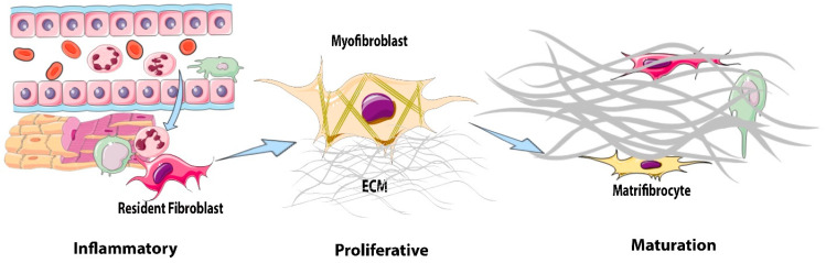
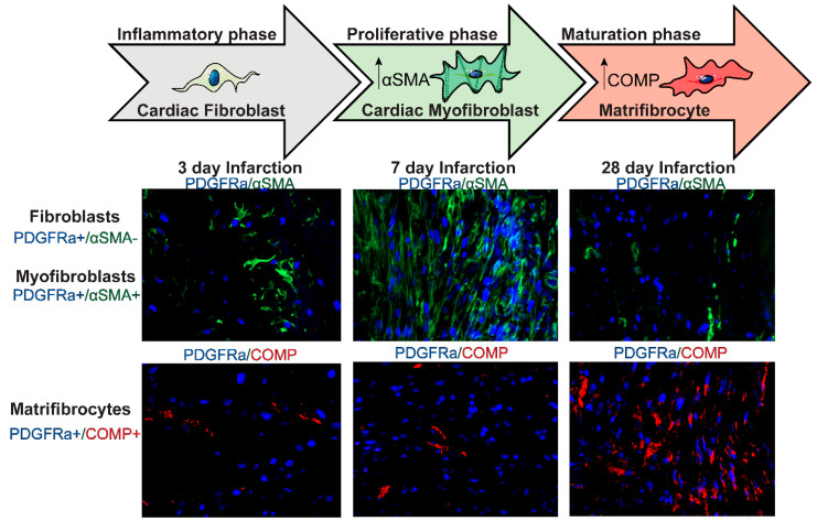
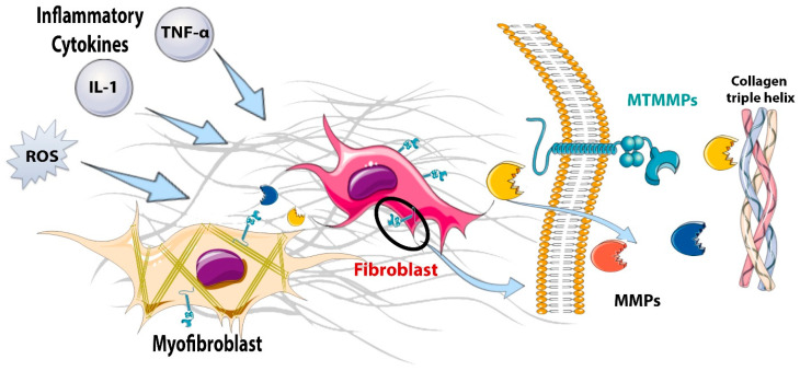
The adult mammalian heart contains abundant interstitial and perivascular fibroblasts that expand following injury and play a reparative role but also contribute to maladaptive fibrotic remodeling. Following myocardial infarction, cardiac fibroblasts undergo dynamic phenotypic transitions, contributing to the regulation of inflammatory, reparative, and angiogenic responses. This review manuscript discusses the mechanisms of regulation, roles and fate of fibroblasts in the infarcted heart. During the inflammatory phase of infarct healing, the release of alarmins by necrotic cells promotes a pro-inflammatory and matrix-degrading fibroblast phenotype that may contribute to leukocyte recruitment. The clearance of dead cells and matrix debris from the infarct stimulates anti-inflammatory pathways and activates transforming growth factor (TGF)-β cascades, resulting in the conversion of fibroblasts to α-smooth muscle actin (α-SMA)-expressing myofibroblasts. Activated myofibroblasts secrete large amounts of matrix proteins and form a collagen-based scar that protects the infarcted ventricle from catastrophic complications, such as cardiac rupture. Moreover, infarct fibroblasts may also contribute to cardiac repair by stimulating angiogenesis. During scar maturation, fibroblasts disassemble α-SMA+ stress fibers and convert to specialized cells that may serve in scar maintenance. The prolonged activation of fibroblasts and myofibroblasts in the infarct border zone and in the remote remodeling myocardium may contribute to adverse remodeling and to the pathogenesis of heart failure. In addition to their phenotypic plasticity, fibroblasts exhibit remarkable heterogeneity. Subsets with distinct phenotypic profiles may be responsible for the wide range of functions of fibroblast populations in infarcted and remodeling hearts.
成年哺乳动物心脏含有丰富的间质和血管周围成纤维细胞,这些细胞在损伤后会扩张,并发挥修复作用,但也会导致适应性不良的纤维化重塑。心肌梗死后,心脏成纤维细胞经历动态的表型转变,有助于调节炎症、修复和血管生成反应。这篇综述讨论了调节机制、成纤维细胞在梗死心脏中的作用和命运。在梗死愈合的炎症阶段,坏死细胞释放警报素促进了促炎和基质降解的成纤维细胞表型,可能有助于白细胞募集。死细胞和基质碎片从梗死区的清除会刺激抗炎途径,并激活转化生长因子 (TGF)-β级联反应,导致成纤维细胞转化为表达α-平滑肌肌动蛋白 (α-SMA)的肌成纤维细胞。活化的肌成纤维细胞大量分泌基质蛋白并形成胶原为基础的瘢痕,保护梗死的心室免受灾难性并发症,如心脏破裂。此外,梗死成纤维细胞也可以通过刺激血管生成来促进心脏修复。在瘢痕成熟过程中,成纤维细胞分解α-SMA+应激纤维,并转化为可能在瘢痕维持中发挥作用的特化细胞。梗死边界区和成纤维细胞重塑心肌中成纤维细胞的长期激活可能导致不良重塑,并导致心力衰竭的发病机制。除了表型可塑性外,成纤维细胞还表现出显著的异质性。具有不同表型特征的亚群可能负责成纤维细胞群体在梗死和重塑心脏中的广泛功能。