Department of Veterinary Medicine (DIMEVET), Università degli Studi di Milano, Via dell'Università 6, 26900, Lodi, Italy.
Centro Ricerca E. Menni, Fondazione Poliambulanza di Brescia, Via Bissolati 57, 25124, Brescia, Italy.
Stem Cell Res Ther. 2020 Mar 4;11(1):99. doi: 10.1186/s13287-020-01611-z.
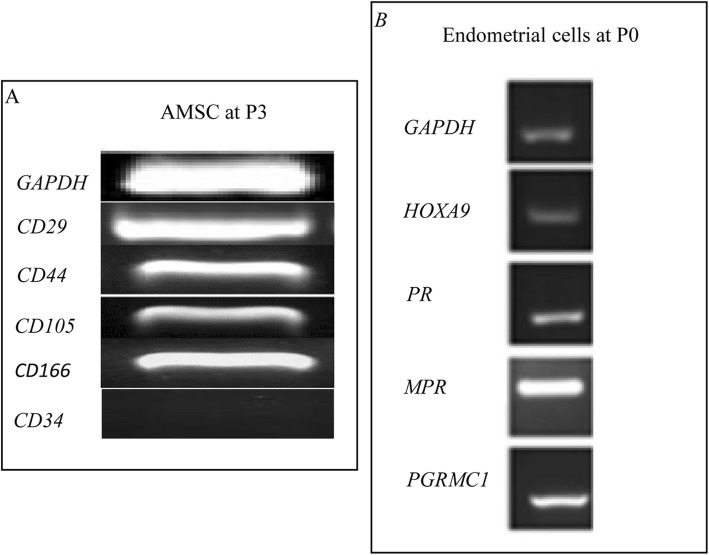
Equine amniotic mesenchymal stromal cells (AMSCs) and their conditioned medium (CM) were evaluated for their ability to inhibit in vitro proliferation of peripheral blood mononuclear cells (PBMCs) with and without priming. Additionally, AMSC immunogenicity was assessed by expression of MHCI and MHCII and their ability to counteract the in vitro inflammatory process.
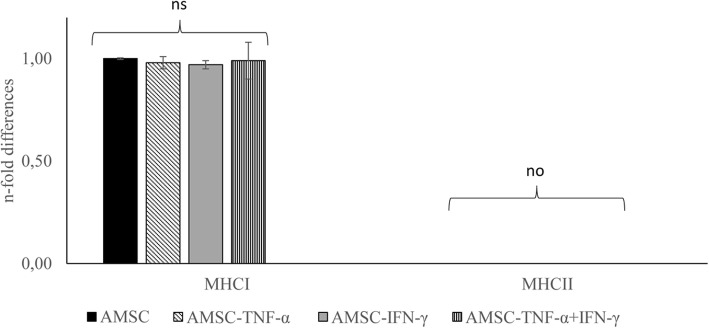
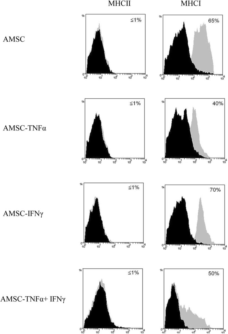
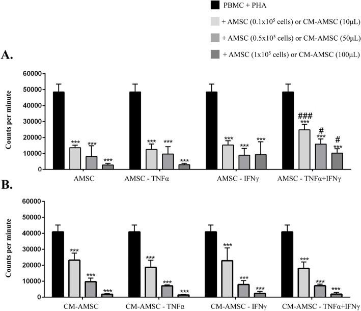
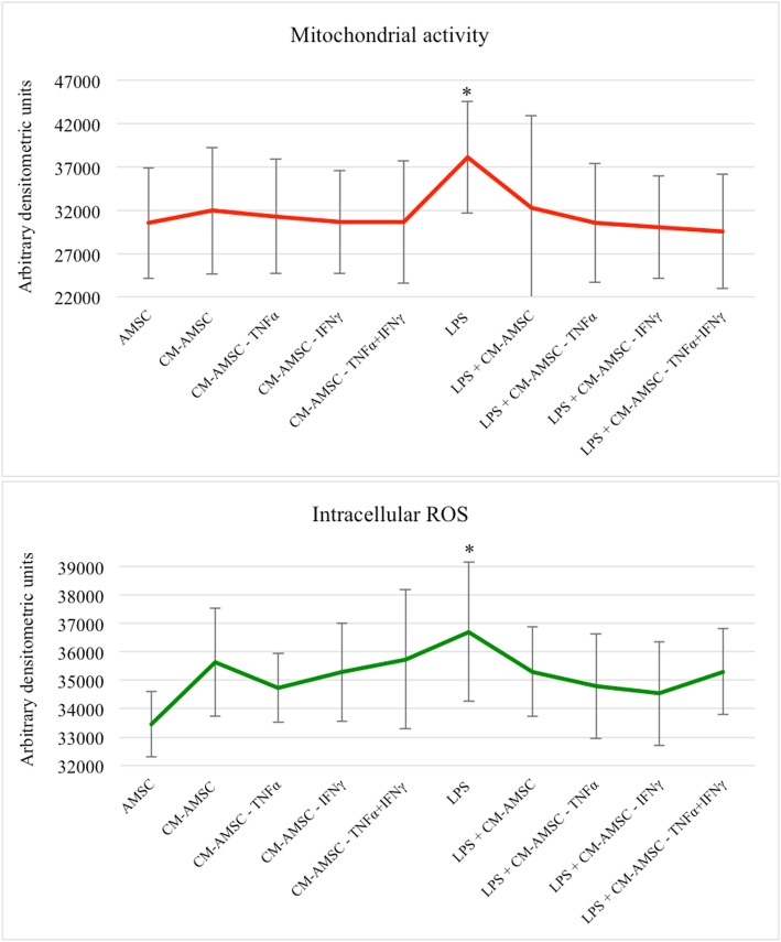
Horse PBMC proliferation was induced with phytohemagglutinin. AMSC priming was performed with 10 ng/ml of TNF-α, 100 ng/ml of IFN-γ, and a combination of 5 ng/ml of TNF-α and 50 ng/ml of IFN-γ. The CM generated from naïve unprimed and primed AMSCs was also tested to evaluate its effects on equine endometrial cells in an in vitro inflammatory model induced by LPS. Immunogenicity marker expression (MHCI and II) was evaluated by qRT-PCR and by flow cytometry.
Priming does not increase MHCI and II expression. Furthermore, the inhibition of PBMC proliferation was comparable between naïve and conditioned cells, with the exception of AMSCs primed with both TNF-α and IFN-γ that had a reduced capacity to inhibit T cell proliferation. However, AMSC viability was lower after priming than under other experimental conditions. CM from naïve and primed AMSCs strongly inhibited PBMC proliferation and counteracted the inflammatory process, rescuing about 65% of endometrial cells treated by LPS.
AMSCs and their CM have a strong capacity to inhibit PBMC proliferation, and priming is not necessary to improve their immunosuppressive activity or reactivity in an inflammatory in vitro model.
评估了马羊膜间充质基质细胞(AMSCs)及其条件培养基(CM)抑制外周血单个核细胞(PBMCs)体外增殖的能力,包括有无预刺激的情况。此外,通过 MHC I 和 MHC II 的表达及其在体外炎症过程中的拮抗能力来评估 AMSC 的免疫原性。
用植物血球凝集素诱导马 PBMC 增殖。用 10ng/ml TNF-α、100ng/ml IFN-γ和 5ng/ml TNF-α和 50ng/ml IFN-γ的组合对 AMSC 进行预刺激。还测试了来自幼稚未刺激和刺激的 AMSC 的 CM,以评估其在 LPS 诱导的体外炎症模型中对马子宫内膜细胞的影响。通过 qRT-PCR 和流式细胞术评估免疫原性标志物(MHC I 和 II)的表达。
预刺激不会增加 MHC I 和 II 的表达。此外,幼稚细胞和条件细胞对 PBMC 增殖的抑制作用相当,除了同时用 TNF-α和 IFN-γ刺激的 AMSCs 抑制 T 细胞增殖的能力降低。然而,与其他实验条件相比,刺激后的 AMSC 活力较低。幼稚和刺激的 AMSC 的 CM 强烈抑制 PBMC 增殖并拮抗炎症过程,挽救了约 65%用 LPS 处理的子宫内膜细胞。
AMSCs 及其 CM 具有强烈抑制 PBMC 增殖的能力,并且预刺激对于提高其在炎症体外模型中的免疫抑制活性或反应性不是必需的。