Guangdong Provincial Key Laboratory of Insect Developmental Biology and Applied Technology, Institute of Insect Science and Technology, School of Life Sciences, South China Normal University, Guangzhou, 510631, China.
Guangzhou Key Laboratory of Insect Development Regulation and Application Research, Institute of Insect Science and Technology, School of Life Sciences, South China Normal University, Guangzhou, 510631, China.
Epigenetics Chromatin. 2020 Mar 5;13(1):12. doi: 10.1186/s13072-020-00334-y.
A large number of in vitro experiments have confirmed that DNA molecules can form i-motif advanced structure when multiple cytosines exist in the sequence. However, whether these structures are present in vivo environment still lacks sufficient experimental evidence.
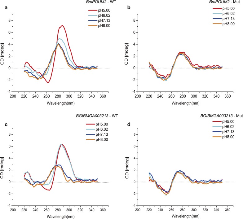
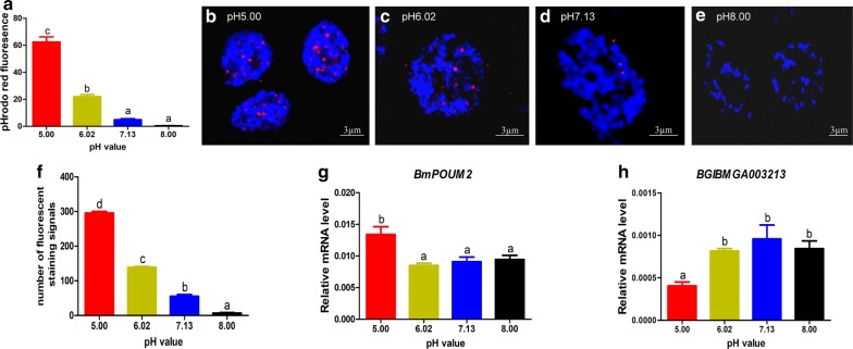
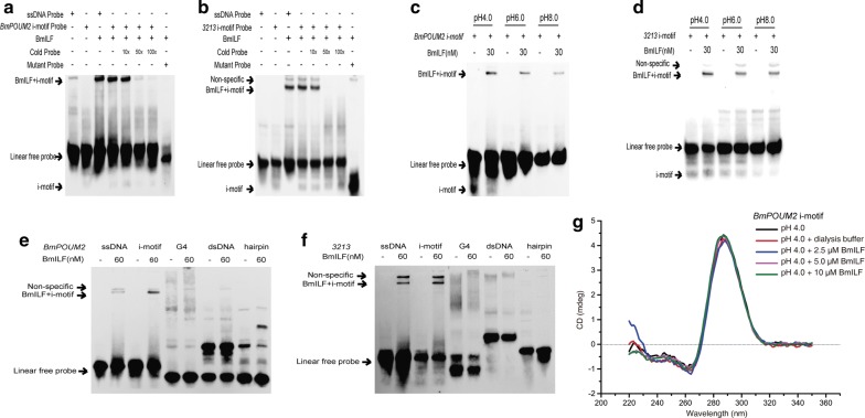
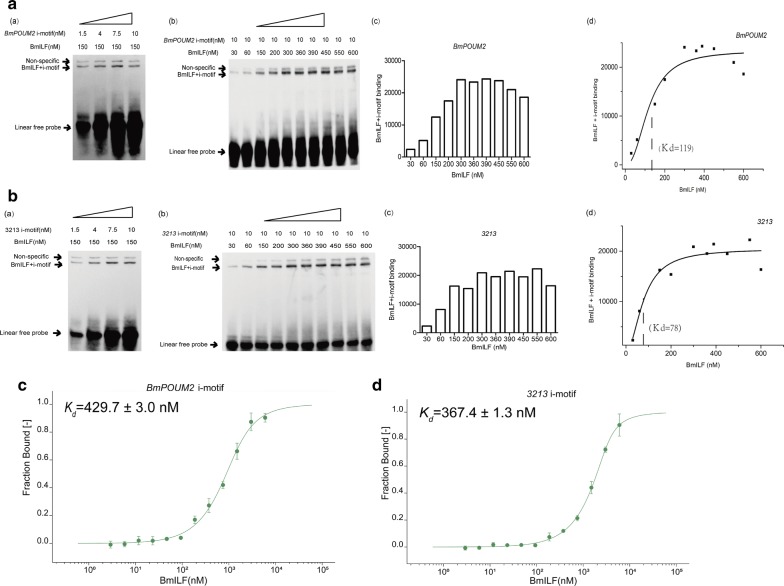
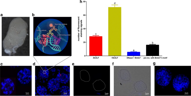
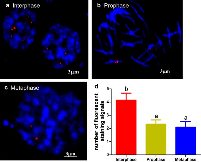
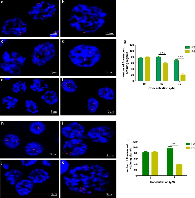
In this paper, we report the in vivo visualization of i-motif structures in the nuclei and chromosomes of the testis of the invertebrate Bombyx mori using immunofluorescence staining with an antibody specifically recognizing the endogenous transcription factor BmILF, which binds i-motif structure with high specificity. The number of i-motif structures observed in the genome increased when the pH was changed from basic to acidic and decreased under treatment with an i-motif inhibitor, the porphyrin compound TMPyP4. The pH change affected the transcription of genes that contain i-motif sequences. Moreover, there were more i-motif structures observed in the testis cells in interphase than in any other cell cycle stage.
In this study, the i-motif structures in invertebrates were detected for the first time at the cell and organ levels. The formation of the structures depended on cell cycle and pH and affected gene expression.
大量体外实验证实,当序列中存在多个胞嘧啶时,DNA 分子可以形成 i- 型发夹结构。然而,这些结构是否存在于体内环境中还缺乏足够的实验证据。
本文通过免疫荧光染色,使用特异性识别内源性转录因子 BmILF 的抗体,报告了在无脊椎动物家蚕的睾丸核和染色体中 i- 型结构的体内可视化。该抗体可以与 i- 型结构高特异性结合。当 pH 值从碱性变为酸性时,观察到基因组中的 i- 型结构数量增加,而在用 i- 型抑制剂卟啉化合物 TMPyP4 处理时则减少。pH 值变化影响了含有 i- 型序列的基因的转录。此外,在有丝分裂期观察到的 i- 型结构比在任何其他细胞周期阶段都多。
本研究首次在细胞和器官水平上检测到了无脊椎动物中的 i- 型结构。结构的形成取决于细胞周期和 pH 值,并影响基因表达。