• 文献检索
  • 文档翻译
  • 深度研究
  • 学术资讯
  • Suppr Zotero 插件Zotero 插件
  • 邀请有礼
  • 套餐&价格
  • 历史记录
应用&插件
Suppr Zotero 插件Zotero 插件浏览器插件Mac 客户端Windows 客户端微信小程序
定价
高级版会员购买积分包购买API积分包
服务
文献检索文档翻译深度研究API 文档MCP 服务
关于我们
关于 Suppr公司介绍联系我们用户协议隐私条款
关注我们

Suppr 超能文献

核心技术专利:CN118964589B侵权必究
粤ICP备2023148730 号-1Suppr @ 2026

文献检索

告别复杂PubMed语法,用中文像聊天一样搜索,搜遍4000万医学文献。AI智能推荐,让科研检索更轻松。

立即免费搜索

文件翻译

保留排版,准确专业,支持PDF/Word/PPT等文件格式,支持 12+语言互译。

免费翻译文档

深度研究

AI帮你快速写综述,25分钟生成高质量综述,智能提取关键信息,辅助科研写作。

立即免费体验

超声造影下乳腺非肿块样病变:特征分析、乳腺影像报告和数据系统分类评估

Breast non-mass-like lesions on contrast-enhanced ultrasonography: Feature analysis, breast image reporting and data system classification assessment.

作者信息

Xu Ping, Yang Min, Liu Yong, Li Yan-Ping, Zhang Hong, Shao Guang-Rui

机构信息

School of Medicine, Shandong University, Jinan 250100, Shandong Province, China.

Department of Ultrasound, Beijing Shijitan Hospital affiliated to Capital Medical University, Beijing 100038, China.

出版信息

World J Clin Cases. 2020 Feb 26;8(4):700-712. doi: 10.12998/wjcc.v8.i4.700.

DOI:10.12998/wjcc.v8.i4.700
PMID:32149054
原文链接:https://pmc.ncbi.nlm.nih.gov/articles/PMC7052556/
Abstract

BACKGROUND

Breast non-mass-like lesions (NMLs) account for 9.2% of all breast lesions. The specificity of the ultrasound diagnosis of NMLs is low, and it cannot be objectively classified according to the 5 Edition of the Breast Imaging Reporting and Data System (BI-RADS). Contrast-enhanced ultrasound (CEUS) can help to differentiate and classify breast lesions but there are few studies on NMLs alone.

AIM

To analyze the features of benign and malignant breast NMLs in grayscale ultrasonography (US), color Doppler flow imaging (CDFI) and CEUS, and to explore the efficacy of the combined diagnosis of NMLs and the effect of CEUS on the BI-RADS classification of NMLs.

METHODS

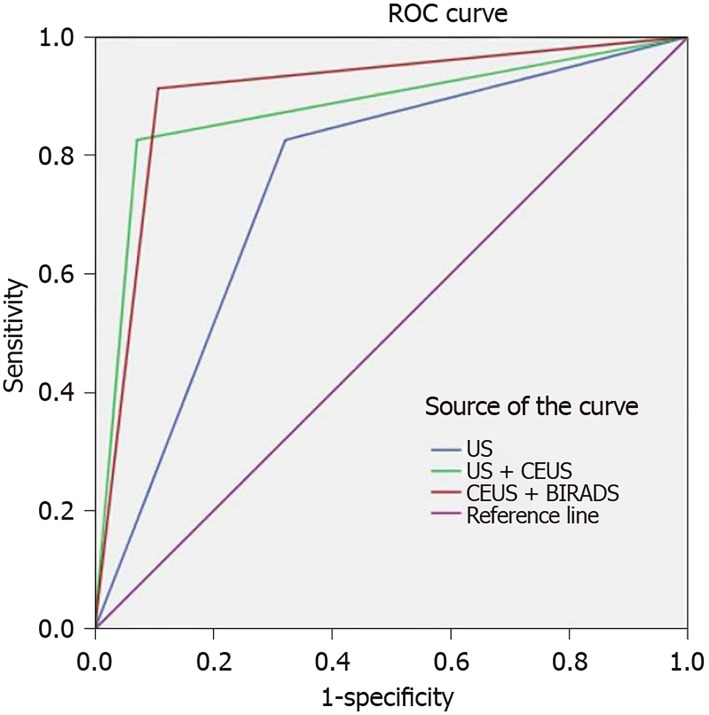
A total of 51 breast NMLs verified by pathology were analyzed in our hospital from January 2017 to April 2019. All lesions were examined by US, CDFI and CEUS, and their features from those examinations were analyzed. With pathology as the gold standard, binary logic regression was used to analyze the independent risk factors for malignant breast NMLs, and a regression equation was established to calculate the efficiency of combined diagnosis. Based on the regression equation, the combined diagnostic efficiency of US combined with CEUS (US + CEUS) was determined. The initial BI-RADS-US classification of NMLs was adjusted according to the independent risk factors identified by CEUS, and the diagnostic efficiency of CEUS combined with BI-RADS (CEUS + BI-RADS) was calculated based on the results. ROC curves were drawn to compare the diagnostic values of the three methods, including US, US + CEUS, and CEUS + BI-RADS, for benign and malignant NMLs.

RESULTS

Microcalcification, enhancement time, enhancement intensity, lesion scope, and peripheral blood vessels were significantly different between benign and malignant NMLs. Among these features, microcalcification, higher enhancement, and lesion scope were identified as independent risk factors for malignant breast NMLs. When US, US + CEUS, and CEUS + BI-RADS were used to identify the benign and malignant breast NMLs, their sensitivity rates were 82.6%, 91.3%, and 87.0%, respectively; their specificity rates were 71.4%, 89.2%, and 92.9%, respectively; their positive predictive values were 70.4%, 87.5%, and 90.9%, respectively; their negative predictive values were 83.3%, 92.6%, and 89.7%, respectively; their accuracy rates were 76.5%, 90.2%, and 90.2%, respectively; and their corresponding areas under ROC curves were 0.752, 0.877 and 0.903, respectively. tests showed that the area under the ROC curve of US was statistically smaller than that of US + CEUS and CEUS + BI-RADS, and there was no statistical difference between US + CEUS and CEUS + BI-RADS.

CONCLUSION

US combined with CEUS can improve diagnostic efficiency for NMLs. The adjustment of the BI-RADS classification according to the features of contrast-enhanced US of NMLs enables the diagnostic results to be simple and intuitive, facilitates the management of NMLs, and effectively reduces the incidence of unnecessary biopsy.

https://cdn.ncbi.nlm.nih.gov/pmc/blobs/6294/7052556/12c7721b5a2d/WJCC-8-700-g004.jpg
https://cdn.ncbi.nlm.nih.gov/pmc/blobs/6294/7052556/4244e7780eae/WJCC-8-700-g001.jpg
https://cdn.ncbi.nlm.nih.gov/pmc/blobs/6294/7052556/0747134fdd3e/WJCC-8-700-g002.jpg
https://cdn.ncbi.nlm.nih.gov/pmc/blobs/6294/7052556/e8f2c2ea7c3f/WJCC-8-700-g003.jpg
https://cdn.ncbi.nlm.nih.gov/pmc/blobs/6294/7052556/12c7721b5a2d/WJCC-8-700-g004.jpg
摘要

背景

乳腺非肿块样病变(NMLs)占所有乳腺病变的9.2%。NMLs的超声诊断特异性较低,且无法根据第5版乳腺影像报告和数据系统(BI-RADS)进行客观分类。超声造影(CEUS)有助于乳腺病变的鉴别和分类,但单独针对NMLs的研究较少。

目的

分析乳腺NMLs在灰阶超声(US)、彩色多普勒血流成像(CDFI)及CEUS中的良恶性特征,探讨NMLs联合诊断的效能以及CEUS对NMLs的BI-RADS分类的影响。

方法

回顾性分析2017年1月至2019年4月我院经病理证实的51例乳腺NMLs。所有病变均行US、CDFI及CEUS检查,并分析其检查特征。以病理为金标准,采用二元逻辑回归分析乳腺NMLs恶性的独立危险因素,建立回归方程计算联合诊断效能。根据回归方程确定US联合CEUS(US + CEUS)的联合诊断效能。根据CEUS确定的独立危险因素调整NMLs的初始BI-RADS-US分类,并根据结果计算CEUS联合BI-RADS(CEUS + BI-RADS)的诊断效能。绘制ROC曲线比较US、US + CEUS及CEUS + BI-RADS三种方法对NMLs良恶性的诊断价值。

结果

乳腺NMLs的微钙化、增强时间、增强强度、病变范围及周边血管在良恶性之间存在显著差异。其中,微钙化、高增强及病变范围被确定为乳腺NMLs恶性的独立危险因素。采用US、US + CEUS及CEUS + BI-RADS鉴别乳腺NMLs的良恶性时,其灵敏度分别为82.6%、91.3%及87.0%;特异度分别为71.4%、89.2%及92.9%;阳性预测值分别为70.4%、87.5%及90.9%;阴性预测值分别为83.3%、92.6%及89.7%;准确率分别为76.5%、90.2%及90.2%;相应的ROC曲线下面积分别为0.752、0.877及0.903。检验显示,US的ROC曲线下面积在统计学上小于US + CEUS及CEUS + BI-RADS,且US + CEUS与CEUS + BI-RADS之间无统计学差异。

结论

US联合CEUS可提高NMLs的诊断效能。根据NMLs的超声造影特征调整BI-RADS分类,使诊断结果简单直观,便于NMLs的管理,有效减少不必要活检的发生率。

相似文献

1
Breast non-mass-like lesions on contrast-enhanced ultrasonography: Feature analysis, breast image reporting and data system classification assessment.超声造影下乳腺非肿块样病变:特征分析、乳腺影像报告和数据系统分类评估
World J Clin Cases. 2020 Feb 26;8(4):700-712. doi: 10.12998/wjcc.v8.i4.700.
2
The role of contrast-enhanced ultrasound in the diagnosis of malignant non-mass breast lesions and exploration of diagnostic criteria.超声造影在诊断恶性非肿块型乳腺病变中的作用及诊断标准探讨。
Br J Radiol. 2021 Apr 1;94(1120):20200880. doi: 10.1259/bjr.20200880. Epub 2021 Feb 9.
3
The kinetic quantitative characteristics of non-mass breast lesions with contrast-enhanced ultrasound: a prospective study.超声造影对非肿块型乳腺病变的定量血流动力学特征研究:前瞻性研究。
Br J Radiol. 2023 Dec;96(1152):20221002. doi: 10.1259/bjr.20221002. Epub 2023 Oct 24.
4
[Diagnostic value of contrast-enhanced ultrasound in breast lesions of BI-RADS 4].超声造影在BI-RADS 4类乳腺病变中的诊断价值
Zhonghua Yi Xue Za Zhi. 2018 May 22;98(19):1498-1502. doi: 10.3760/cma.j.issn.0376-2491.2018.19.009.
5
Application of contrast-enhanced ultrasound in the diagnosis of small breast lesions.超声造影在乳腺小病灶诊断中的应用
Clin Hemorheol Microcirc. 2018;70(3):291-300. doi: 10.3233/CH-170368.
6
BI-RADS 4 breast lesions: could multi-mode ultrasound be helpful for their diagnosis?乳腺影像报告和数据系统(BI-RADS)4类乳腺病变:多模式超声对其诊断有帮助吗?
Gland Surg. 2019 Jun;8(3):258-270. doi: 10.21037/gs.2019.05.01.
7
Ultrasound classification of non-mass breast lesions following BI-RADS presents high positive predictive value.超声 BI-RADS 分类对非肿块性乳腺病变具有较高的阳性预测值。
PLoS One. 2022 Nov 30;17(11):e0278299. doi: 10.1371/journal.pone.0278299. eCollection 2022.
8
Predictive value of contrast-enhanced ultrasonography and ultrasound elastography for management of BI-RADS category 4 nonpalpable breast masses.超声造影及超声弹性成像对 BI-RADS 分类 4 类触诊阴性乳腺肿块的预测价值。
Eur J Radiol. 2024 Apr;173:111391. doi: 10.1016/j.ejrad.2024.111391. Epub 2024 Feb 28.
9
Diagnosis of sub-centimetre breast lesions: combining BI-RADS-US with strain elastography and contrast-enhanced ultrasound-a preliminary study in China.乳腺亚厘米级病变的诊断:联合乳腺影像报告和数据系统-超声(BI-RADS-US)、应变弹性成像及超声造影在中国的初步研究
Eur Radiol. 2017 Jun;27(6):2443-2450. doi: 10.1007/s00330-016-4628-4. Epub 2016 Oct 19.
10
The diagnostic value of contrast-enhanced ultrasonography in breast ductal abnormalities.增强超声在乳腺导管异常中的诊断价值。
Cancer Imaging. 2023 Mar 10;23(1):25. doi: 10.1186/s40644-023-00539-w.

引用本文的文献

1
Incremental Value of Shear Wave Elastography and Contrast-Enhanced Ultrasound in the Differential Diagnosis of Breast Non-Mass-Like Lesions.剪切波弹性成像和超声造影在乳腺非肿块样病变鉴别诊断中的增量价值
Int J Womens Health. 2024 Dec 20;16:2221-2230. doi: 10.2147/IJWH.S490565. eCollection 2024.
2
Diagnostic value of multimodal ultrasound for breast cancer and prediction of sentinel lymph node metastases.多模态超声对乳腺癌的诊断价值及前哨淋巴结转移的预测
Front Cell Dev Biol. 2024 Sep 5;12:1431883. doi: 10.3389/fcell.2024.1431883. eCollection 2024.
3
Current status and future perspectives of contrast-enhanced ultrasound diagnosis of breast lesions.

本文引用的文献

1
Non-Mass Breast Lesions on Ultrasound: Feature Exploration and Multimode Ultrasonic Diagnosis.超声检查发现的非肿块性乳腺病变:特征探索与多模态超声诊断
Ultrasound Med Biol. 2018 Aug;44(8):1703-1711. doi: 10.1016/j.ultrasmedbio.2018.05.005. Epub 2018 May 31.
2
Association between enhancement patterns and parameters of contrast-enhanced ultrasound and microvessel distribution in breast cancer.超声造影增强模式及参数与乳腺癌微血管分布的相关性
Oncol Lett. 2018 Apr;15(4):5643-5649. doi: 10.3892/ol.2018.8078. Epub 2018 Feb 16.
3
B-Mode Ultrasound Combined with Color Doppler and Strain Elastography in the Diagnosis of Non-mass Breast Lesions: A Prospective Study.
乳腺病变超声造影诊断的现状与展望。
J Med Ultrason (2001). 2024 Oct;51(4):611-625. doi: 10.1007/s10396-024-01486-0. Epub 2024 Aug 23.
4
Determining whether the diagnostic value of B-ultrasound combined with contrast-enhanced ultrasound and shear wave elastography in breast mass-like and non-mass-like lesions differs: a diagnostic test.确定B超联合超声造影及剪切波弹性成像在乳腺肿块样和非肿块样病变中的诊断价值是否存在差异:一项诊断试验。
Gland Surg. 2023 Feb 28;12(2):282-296. doi: 10.21037/gs-23-51. Epub 2023 Feb 24.
5
Non-mass Breast Lesions: Could Multimodal Ultrasound Imaging Be Helpful for Their Diagnosis?非肿块性乳腺病变:多模态超声成像对其诊断有帮助吗?
Diagnostics (Basel). 2022 Nov 23;12(12):2923. doi: 10.3390/diagnostics12122923.
6
Development and internal validation of a conventional ultrasound-based nomogram for predicting malignant nonmasslike breast lesions.用于预测乳腺非肿块样恶性病变的传统超声列线图的开发与内部验证
Quant Imaging Med Surg. 2022 Dec;12(12):5452-5461. doi: 10.21037/qims-22-378.
7
Sports Medical Image Modeling of Injury Prevention in Wushu Training.武术训练中预防损伤的运动医学影像建模。
Scanning. 2022 Aug 9;2022:5201952. doi: 10.1155/2022/5201952. eCollection 2022.
8
The clinical value of conventional ultrasound combined with contrast-enhanced ultrasound in the evaluation of BI-RADS 4 lesions detected by magnetic resonance imaging.常规超声联合超声造影在磁共振成像检测 BI-RADS 4 病变评估中的临床价值。
Br J Radiol. 2022 Aug 1;95(1136):20220025. doi: 10.1259/bjr.20220025. Epub 2022 May 30.
9
The role of contrast-enhanced ultrasound in the diagnosis of malignant non-mass breast lesions and exploration of diagnostic criteria.超声造影在诊断恶性非肿块型乳腺病变中的作用及诊断标准探讨。
Br J Radiol. 2021 Apr 1;94(1120):20200880. doi: 10.1259/bjr.20200880. Epub 2021 Feb 9.
10
Does contrast-enhanced ultrasound (CEUS) play a better role in diagnosis of breast lesions with calcification? A comparison with MRI.超声造影(CEUS)在钙化性乳腺病变的诊断中是否发挥着更好的作用?与磁共振成像(MRI)的比较。
Br J Radiol. 2020 Aug;93(1112):20200195. doi: 10.1259/bjr.20200195. Epub 2020 Jun 2.
B超联合彩色多普勒及应变弹性成像在非肿块型乳腺病变诊断中的应用:一项前瞻性研究
Ultrasound Med Biol. 2017 Nov;43(11):2582-2590. doi: 10.1016/j.ultrasmedbio.2017.07.014. Epub 2017 Aug 26.
4
The role of microvessel density, lymph node metastasis, and tumor size as prognostic factors of distant metastasis in colorectal cancer.微血管密度、淋巴结转移及肿瘤大小作为结直肠癌远处转移预后因素的作用。
Oncol Lett. 2017 Jun;13(6):4327-4333. doi: 10.3892/ol.2017.5959. Epub 2017 Mar 31.
5
Clinical Implications for Germline PTEN Spectrum Disorders.种系 PTEN 谱疾病的临床意义。
Endocrinol Metab Clin North Am. 2017 Jun;46(2):503-517. doi: 10.1016/j.ecl.2017.01.013. Epub 2017 Mar 18.
6
Non-mass breast lesions on ultrasound: final outcomes and predictors of malignancy.超声检查发现的非肿块性乳腺病变:最终结局及恶性肿瘤预测因素
Acta Radiol. 2017 Sep;58(9):1054-1060. doi: 10.1177/0284185116683574. Epub 2017 Jan 23.
7
Non-mass lesions on screening breast ultrasound.乳腺超声筛查中的非肿块性病变
Med Ultrason. 2016 Dec 5;18(4):446-451. doi: 10.11152/mu-871.
8
Genomic Changes in Normal Breast Tissue in Women at Normal Risk or at High Risk for Breast Cancer.正常风险或高风险乳腺癌女性正常乳腺组织中的基因组变化。
Breast Cancer (Auckl). 2016 Aug 17;10:109-46. doi: 10.4137/BCBCR.S39384. eCollection 2016.
9
Incorporating Contrast-Enhanced Ultrasound into the BI-RADS Scoring System Improves Accuracy in Breast Tumor Diagnosis: A Preliminary Study in China.将超声造影纳入BI-RADS评分系统可提高乳腺肿瘤诊断的准确性:一项中国的初步研究。
Ultrasound Med Biol. 2016 Nov;42(11):2630-2638. doi: 10.1016/j.ultrasmedbio.2016.07.005. Epub 2016 Aug 17.
10
Additional diagnostic value of shear-wave elastography and color Doppler US for evaluation of breast non-mass lesions detected at B-mode US.剪切波弹性成像和彩色多普勒超声对B超检测出的乳腺非肿块性病变评估的附加诊断价值。
Eur Radiol. 2016 Oct;26(10):3542-9. doi: 10.1007/s00330-015-4201-6. Epub 2016 Jan 19.