Institute of Psychiatry, Psychology and Neuroscience, King's College London, Department of Psychological Medicine, London, UK.
National Institute for Health and Research Biomedical Research Centre at South London and Maudsley NHS Foundation Trust and King's College London, London, UK.
Transl Psychiatry. 2020 Mar 9;10(1):89. doi: 10.1038/s41398-020-0768-z.
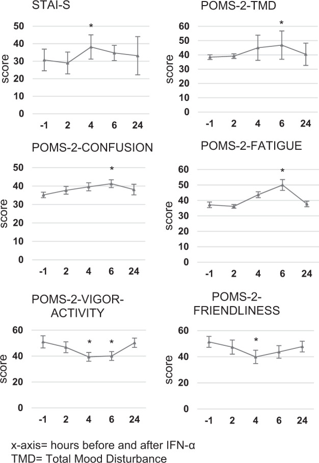
Depression is associated with peripheral inflammation, but its link with brain microglial activity remains unclear. In seven healthy males, we used repeated translocator protein-Positron Emission Tomography (TSPO-PET) dynamic scans with [C]PBR28 to image brain microglial activation before and 24 h after the immune challenge interferon (IFN)-α. We also investigated the association between changes in peripheral inflammation, changes in microglial activity, and changes in mood. IFN-α administration decreased [C]PBR28 PET tissue volume of distribution (Vt) across the brain (-20 ± 4%; t = 4.1, p = 0.01), but after correction for radioligand free-plasma fraction there were no longer any changes (+23 ± 31%; t = 0.1, p = 0.91). IFN-α increased serum IL-6 (1826 ± 513%, t = -7.5, p < 0.001), IL-7 (39 ± 12%, t = -3.6, p = 0.01), IL-10 (328 ± 48%, t = -12.8, p < 0.001), and IFN-γ (272 ± 64%, t = -7.0, p < 0.001) at 4-6 h, and increased serum TNF-α (49 ± 7.6%, t = -7.5, p < 0.001), IL-8 (39 ± 12%, t = -3.5, p = 0.013), and C-reactive protein (1320 ± 459%, t = -7.2, p < 0.001) at 24 h. IFN-α induced temporary mood changes and sickness symptoms after 4-6 h, measured as an increase in POMS-2 total mood score, confusion and fatigue, and a decrease in vigor and friendliness (all p ≤ 0.04). No association was found between changes in peripheral inflammation and changes in PET or mood measures. Our work suggests that brain TSPO-PET signal is highly dependent of inflammation-induced changes in ligand binding to plasma proteins. This limits its usefulness as a sensitive marker of neuroinflammation and consequently, data interpretation. Thus, our results can be interpreted as showing either that [C]PBR28 is not sensitive enough under these conditions, or that there is simply no microglial activation in this model.
抑郁症与外周炎症有关,但它与大脑小胶质细胞活性的联系尚不清楚。在 7 名健康男性中,我们使用重复的[11C]PBR28 正电子发射断层扫描(TSPO-PET)动态扫描,在免疫挑战干扰素(IFN)-α之前和之后 24 小时,对大脑小胶质细胞的激活进行成像。我们还研究了外周炎症变化、小胶质细胞活性变化和情绪变化之间的关联。IFN-α 给药降低了整个大脑的[11C]PBR28 PET 组织分布容积(Vt)(-20±4%;t=4.1,p=0.01),但在对配体结合血浆分数进行校正后,不再有任何变化(+23±31%;t=0.1,p=0.91)。IFN-α 增加了血清白细胞介素-6(1826±513%,t=-7.5,p<0.001)、白细胞介素-7(39±12%,t=-3.6,p=0.01)、白细胞介素-10(328±48%,t=-12.8,p<0.001)和 IFN-γ(272±64%,t=-7.0,p<0.001)在 4-6 小时内,同时增加了血清肿瘤坏死因子-α(49±7.6%,t=-7.5,p<0.001)、白细胞介素-8(39±12%,t=-3.5,p=0.013)和 C 反应蛋白(1320±459%,t=-7.2,p<0.001)在 24 小时内。IFN-α 在 4-6 小时后引起暂时的情绪变化和疾病症状,表现为 POMS-2 总情绪评分、困惑和疲劳增加,活力和友好度下降(所有 p≤0.04)。外周炎症变化与 PET 或情绪测量值之间没有发现相关性。我们的工作表明,大脑 TSPO-PET 信号对配体与血浆蛋白结合引起的炎症变化高度依赖。这限制了它作为神经炎症敏感标志物的用途,因此,数据解释也是如此。因此,我们的结果可以解释为,要么在这些条件下 [11C]PBR28 不够敏感,要么在这个模型中根本没有小胶质细胞激活。