College of Animal Science, Zhejiang University, 310058 Hangzhou, China.
Zhejiang Animal Husbandry Techniques Extension Station, 310020 Hangzhou, China.
Cells. 2020 Mar 6;9(3):645. doi: 10.3390/cells9030645.
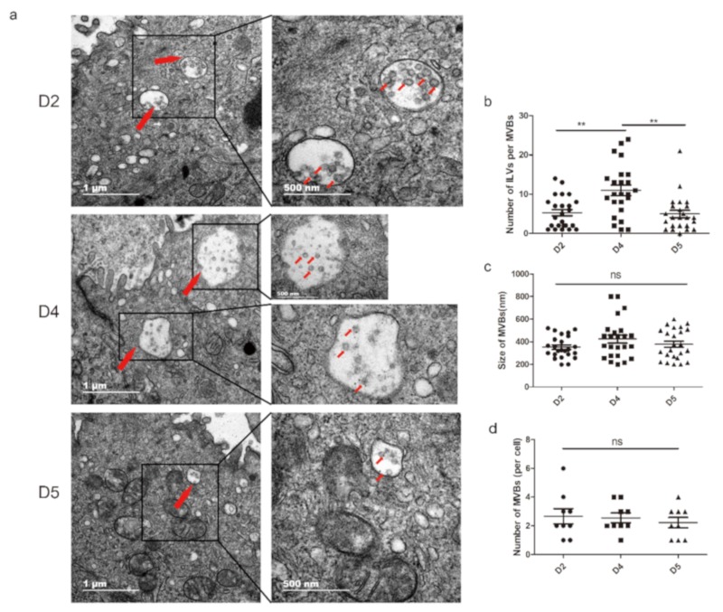
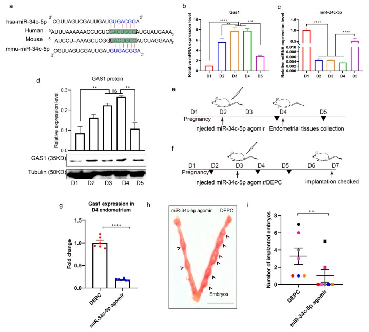
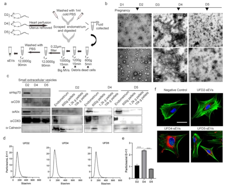
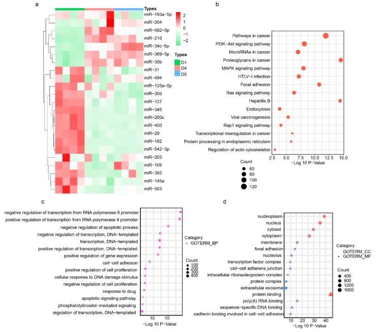
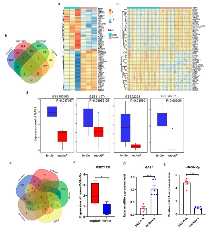
Synchronous communication between the developing embryo and the receptive endometrium is crucial for embryo implantation. Thus, uterine receptivity evaluation is vital in managing recurrent implantation failure (RIF). The potential roles of small extracellular vesicle (sEV) miRNAs in pregnancy have been widely studied. However, the systematic study of sEVs derived from endometrium and its cargos during the implantation stage have not yet been reported. In this study, we isolated endometrium-derived sEVs from the mouse endometrium on D2 (pre-receptive phase), D4 (receptive phase), and D5 (implantation) of pregnancy. Herein, we reveal that multivesicular bodies (MVBs) in the endometrium increase in number during the window of implantation (WOI). Moreover, our findings indicate that CD63, a well-known sEV marker, is expressed in the luminal and glandular epithelium of mouse endometrium. The sEV miRNA expression profiles indicated that miR-34c-5p, miR-210, miR-369-5p, miR-30b, and miR-582-5p are enriched during WOI. Further, we integrated the RIF's database analysis results and found out that miR-34c-5p regulates growth arrest specific 1 (GAS1) for normal embryo implantation. Notably, miR-34c-5p is downregulated during implantation but upregulated in sEVs. An implication of this is the possibility that sEVs miR-34c-5p could be used to evaluate uterine states. In conclusion, these findings suggest that the endometrium derived-sEV miRNAs are potential biomarkers in determining the appropriate period for embryo implantation. This study also has several important implications for future practice, including therapy of infertility.
胚胎发育与接受性子宫内膜之间的同步通讯对胚胎着床至关重要。因此,子宫接受性评估对于管理反复着床失败(RIF)至关重要。小细胞外囊泡(sEV)miRNA 在妊娠中的潜在作用已得到广泛研究。然而,在着床阶段,尚未有关于来源于子宫内膜及其 cargo 的 sEV 的系统研究。在这项研究中,我们从小鼠妊娠的 D2(预接受期)、D4(接受期)和 D5(着床期)子宫内膜中分离出子宫内膜来源的 sEV。在此,我们揭示了在着床窗口期(WOI)期间,子宫内膜中的多泡体(MVB)数量增加。此外,我们的研究结果表明,CD63,一种众所周知的 sEV 标志物,在小鼠子宫内膜的腔上皮和腺上皮中表达。sEV miRNA 表达谱表明,miR-34c-5p、miR-210、miR-369-5p、miR-30b 和 miR-582-5p 在 WOI 期间富集。此外,我们整合了 RIF 的数据库分析结果,发现 miR-34c-5p 调节胚胎着床正常所需的生长停滞特异基因 1(GAS1)。值得注意的是,miR-34c-5p 在着床过程中下调,但在 sEV 中上调。这意味着 sEVs miR-34c-5p 可能被用于评估子宫状态。总之,这些发现表明,子宫内膜来源的 sEV miRNAs 可能是确定胚胎着床适当时期的潜在生物标志物。本研究对未来的实践也有几个重要的启示,包括不孕的治疗。