Thayer School of Engineering at Dartmouth College, Hanover, New Hampshire, United States of America.
Geisel School of Medicine at Dartmouth College, Hanover, New Hampshire, United States of America.
PLoS One. 2020 Mar 11;15(3):e0230267. doi: 10.1371/journal.pone.0230267. eCollection 2020.
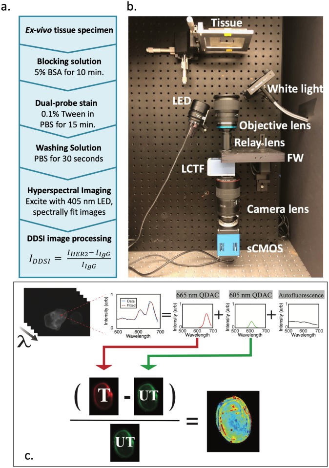
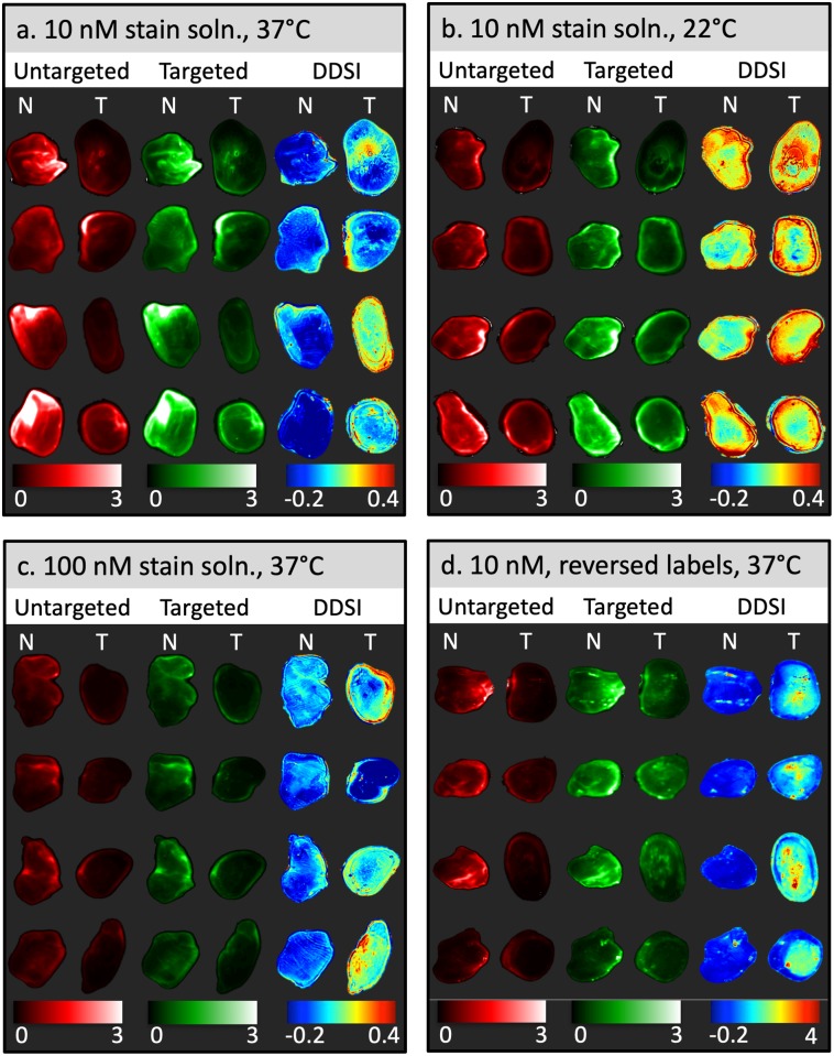
Rapid, intra-operative identification of tumor tissue in the margins of excised specimens has become an important focus in the pursuit of reducing re-excision rates, especially for breast conserving surgery. Dual-probe difference specimen imaging (DDSI) is an emerging approach that uses the difference in uptake/clearance kinetics between a pair of fluorescently-labeled stains, one targeted to a biomarker-of-interest and the other an untargeted isotype, to reveal receptor-specific images of the specimen. Previous studies using antibodies labeled with either enhanced Raman particles or organic fluorophores have shown promising tumor vs. normal diagnostic performance. Yet, the unique properties of quantum dot-labeled antibody complexes (QDACs), which provide spectrally-distinct fluorescence emission from a common excitation source, make them ideal candidates for this application. Herein, we evaluate the diagnostic performance of QDAC-based DDSI in excised xenografts.
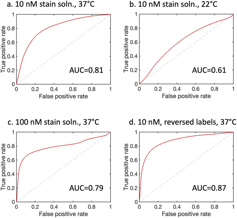
Excised fresh specimens of normal tissue and human tumor xenografts with elevated expression of HER2 were stained with a HER2-targeted QDAC and an untargeted QDAC isotype. Stained specimens were imaged on a custom hyperspectral imaging system capable of spectrally separating the quantum dot signatures, and images processed using the DDSI approach. The diagnostic performance of this technique under different incubation temperatures and probe concentrations was evaluated using receiver-operator characteristic analysis.
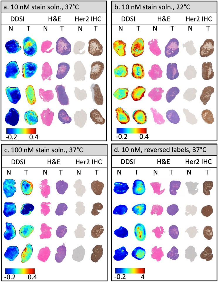
HER2-targeted QDAC-DDSI was able to distinguish HER2(+) tumors from normal tissue with reasonably high diagnostic performance; however, this performance was sensitive to temperature during the staining procedure. Area under the curve values were 0.61 when staining at room temperature but increased to over 0.81 when staining at 37 °C. Diagnostic performance was not affected by increasing stain concentration.
This study is the first to report dual-probe difference imaging of specimens using QDACs and hyperspectral imaging. Our results show promising diagnostic performance under certain conditions, and compel further optimization and evaluation of this intra-operative margin assessment technique.
在切除标本的边缘快速、术中识别肿瘤组织已成为降低再次切除率的重要关注点,尤其是对于保乳手术。双探头差异标本成像(DDSI)是一种新兴方法,它利用一对荧光标记的染料之间摄取/清除动力学的差异,一种针对感兴趣的生物标志物,另一种为非靶向同型,以显示标本的受体特异性图像。使用标记有增强拉曼粒子或有机荧光团的抗体的先前研究表明具有有希望的肿瘤与正常诊断性能。然而,量子点标记抗体复合物(QDAC)的独特特性提供了来自共同激发源的光谱上不同的荧光发射,使其成为该应用的理想候选者。在这里,我们评估基于 QDAC 的 DDSI 在切除异种移植物中的诊断性能。
用 HER2 靶向 QDAC 和非靶向 QDAC 同型对正常组织和人肿瘤异种移植物的新鲜切除标本进行染色。用能够光谱分离量子点特征的定制高光谱成像系统对染色标本进行成像,并使用 DDSI 方法处理图像。使用接收器操作特性分析评估不同孵育温度和探针浓度下该技术的诊断性能。
HER2 靶向 QDAC-DDSI 能够以合理的高诊断性能区分 HER2(+)肿瘤与正常组织;然而,这种性能对染色过程中的温度敏感。在室温下染色时,曲线下面积为 0.61,但在 37°C 时增加到 0.81 以上。增加染色浓度不会影响诊断性能。
这项研究首次报道了使用 QDAC 和高光谱成像进行双探针差异成像。我们的结果表明,在某些条件下具有有希望的诊断性能,并迫使进一步优化和评估这种术中边缘评估技术。