Biomedical Engineering Department, Oregon Health & Science University, 2730 S Moody Ave, Mail Code: CL3SG, Portland, OR, 97201, USA.
Thayer School of Engineering, Dartmouth College, Hanover, NH, 03755, USA.
BMC Cancer. 2021 Apr 21;21(1):440. doi: 10.1186/s12885-021-08179-8.
Re-excision rates following breast conserving surgery (BCS) remain as high as ~ 35%, with positive margins detected during follow-up histopathology. Additional breast cancer resection surgery is not only taxing on the patient and health care system, but also delays adjuvant therapies, increasing morbidity and reducing the likelihood of a positive outcome. The ability to precisely resect and visualize tumor margins in real time within the surgical theater would greatly benefit patients, surgeons and the health care system. Current tumor margin assessment technologies utilized during BCS involve relatively lengthy and labor-intensive protocols, which impede the surgical work flow.
In previous work, we have developed and validated a fluorescence imaging method termed dual probe difference specimen imaging (DDSI) to accurately detect benign and malignant tissue with direct correlation to the targeted biomarker expression levels intraoperatively. The DDSI method is currently on par with touch prep cytology in execution time (~ 15-min). In this study, the main goal was to shorten the DDSI protocol by decreasing tissue blocking and washing times to optimize the DDSI protocol to < 10-min whilst maintaining robust benign and malignant tissue differentiation.
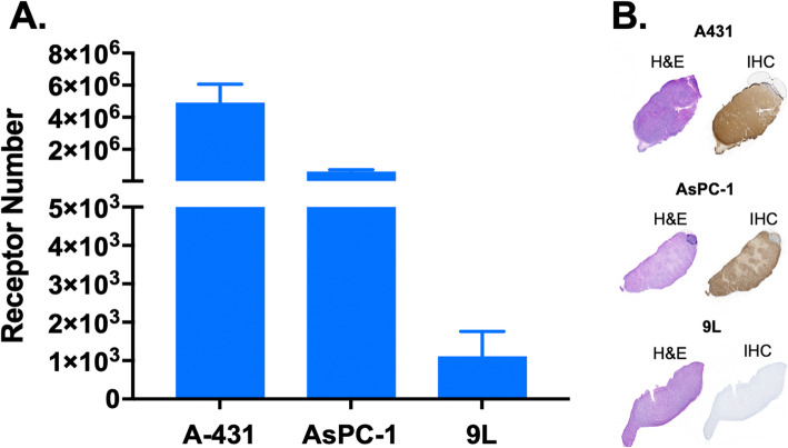
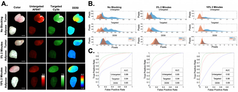
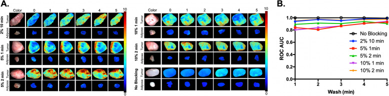
We evaluated the utility of the shortened DDSI staining methodology using xenografts grown from cell lines with varied epidermal growth factor receptor (EGFR) expression levels, comparing accuracy through receiver operator characteristic (ROC) curve analyses across varied tissue blocking and washing times. An optimized 8-min DDSI methodology was developed for future clinical translation.
Successful completion of this work resulted in substantial shortening of the DDSI methodology for use in the operating room, that provided robust, highly receptor specific, sensitive diagnostic capabilities between benign and malignant tissues.
保乳手术后(BCS)的再次切除率仍然高达约 35%,在随访组织病理学检查中发现阳性边缘。额外的乳腺癌切除术不仅对患者和医疗保健系统造成负担,而且还会延迟辅助治疗,增加发病率并降低阳性结果的可能性。在手术室内实时精确切除和可视化肿瘤边缘的能力将极大地造福于患者、外科医生和医疗保健系统。目前在 BCS 中使用的肿瘤边缘评估技术涉及相对冗长和劳动密集型的方案,这会阻碍手术工作流程。
在以前的工作中,我们开发并验证了一种荧光成像方法,称为双探针差异标本成像(DDSI),可以在手术室内准确检测良性和恶性组织,并且与靶向生物标志物表达水平直接相关。DDSI 方法在执行时间上与触摸准备细胞学相当(~15 分钟)。在这项研究中,主要目标是通过减少组织阻断和洗涤时间来缩短 DDSI 方案,以优化 DDSI 方案至<10 分钟,同时保持稳健的良性和恶性组织分化。
我们使用具有不同表皮生长因子受体(EGFR)表达水平的细胞系生长的异种移植物评估了缩短的 DDSI 染色方法的实用性,通过比较在不同组织阻断和洗涤时间下的接收者操作特征(ROC)曲线分析来比较准确性。开发了一种优化的 8 分钟 DDSI 方法,用于未来的临床转化。
这项工作的成功完成导致 DDSI 方法的实质性缩短,可用于手术室,为良性和恶性组织之间提供强大、高度受体特异性、敏感的诊断能力。