Department of Neurosurgery, Keio University School of Medicine, 35 Shinanomachi, Shinjuku-ku, Tokyo, 160-8582, Japan.
Department of Neurosurgery, Hiratsuka City Hospital, Hiratsuka, Kanagawa, 254-0019, Japan.
BMC Cancer. 2020 Mar 12;20(1):196. doi: 10.1186/s12885-020-6589-x.
The expression of vascular endothelial growth factor (VEGF)-A/ VAGF receptors (VEGFRs) signaling plays a pivotal role in the tumor angiogenesis and the development of the immunosuppressive tumor microenvironment in glioblastomas. We have previously conducted exploratory clinical studies investigating VEGFRs peptide vaccination with and without multiple glioma oncoantigens in patients with recurrent high-grade gliomas. Recently, an exploratory clinical investigation of VEGFRs peptide vaccination was conducted in patients with progressive neurofibromatosis type 2. Those studies suggested that cytotoxic T lymphocytes (CTLs) induced by the vaccination can directly kill a wide variety of cells associated with tumor growth, including tumor vessels, tumor cells, and immunosuppressive cells expressing VEGFR1 and/or 2. In the present study, synergistic activity of the combination of VEGFRs peptide vaccination with chemotherapy was evaluated.
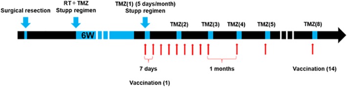
We performed the first clinical trial to assess VEGFR1 and 2 vaccination along with temozolomide (TMZ) -based chemoradiotherapy for the patients with primary glioblastomas. Furthermore, histopathological changes after the vaccination were evaluated using paired pre- and post- vaccination specimens.
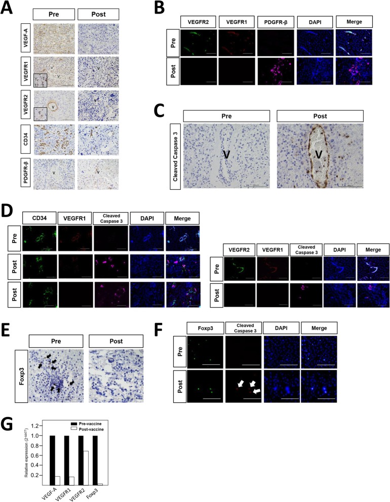
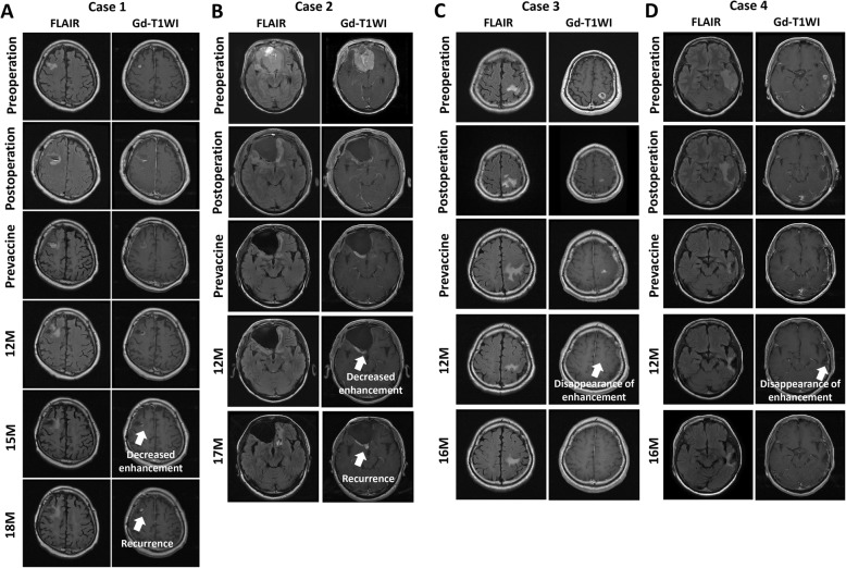
The disappearance of radiographically enhanced lesion was observed in 2 patients after the vaccination, including one in which the methylation of the O6-methylguanine-DNA methyltransferase (MGMT) promoter was not observed. The histopathological findings of pre- and post-vaccination specimens demonstrated that tumor vessels showed negative or slight VEGFRs expressions after the vaccination and most endothelial cells were covered with PDGFR-β-positive pericytes. Notably, CTLs induced by VEGFRs peptide vaccination attacked not only tumor vessels but also tumor cells and regulatory T cells expressing VEGFRs even in recurrent tumors.
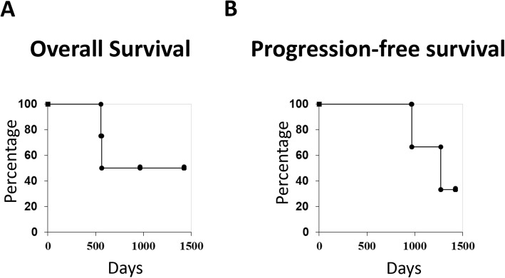
VEGFR1 and 2 vaccination may have a preliminary synergistic effect when administered with TMZ. The limitation of the present study was the paucity of the number of the samples. Further studies involving more patients are warranted to confirm the findings of this study.
This study was registered as UMIN000013381 (University Hospital Medical Information Network-Clinical Trial Registry: UMIN-CTR) on 5 March, 2014 and with the Japan Registry of Clinical Trials (jRCT) as jRCTs031180170 on 1 March, 2019.
血管内皮生长因子(VEGF)-A/VEGF 受体(VEGFR)信号的表达在胶质母细胞瘤的肿瘤血管生成和免疫抑制肿瘤微环境的发展中起着关键作用。我们之前进行了探索性临床研究,调查了在复发性高级别胶质瘤患者中使用和不使用多种神经胶质瘤肿瘤抗原的 VEGFR 肽疫苗接种。最近,对进展性神经纤维瘤病 2 型患者进行了 VEGFR 肽疫苗接种的探索性临床研究。这些研究表明,疫苗接种诱导的细胞毒性 T 淋巴细胞(CTL)可以直接杀死与肿瘤生长相关的多种细胞,包括肿瘤血管、肿瘤细胞和表达 VEGFR1 和/或 2 的免疫抑制细胞。在本研究中,评估了 VEGFR 肽疫苗接种与化疗联合的协同活性。
我们进行了第一项临床试验,以评估原发性胶质母细胞瘤患者接受 VEGFR1 和 2 疫苗接种联合替莫唑胺(TMZ)为基础的放化疗的效果。此外,使用配对的接种前后标本评估接种后的组织病理学变化。
接种后 2 例患者的影像学增强病变消失,其中 1 例 O6-甲基鸟嘌呤-DNA 甲基转移酶(MGMT)启动子的甲基化未观察到。接种前后标本的组织病理学发现表明,接种后肿瘤血管的 VEGFRs 表达呈阴性或轻微,大多数内皮细胞被 PDGFR-β 阳性周细胞覆盖。值得注意的是,VEGFR 肽疫苗接种诱导的 CTL 不仅攻击肿瘤血管,而且攻击表达 VEGFRs 的肿瘤细胞和调节性 T 细胞,即使在复发性肿瘤中也是如此。
VEGFR1 和 2 疫苗接种与 TMZ 联合使用可能具有初步的协同作用。本研究的局限性是样本数量较少。需要进一步的多患者研究来证实本研究的结果。
本研究于 2014 年 3 月 5 日在大学医院医学信息网络-临床试验注册处(UMIN000013381)和日本临床试验注册处(jRCT)以 jRCTs031180170 注册。