Başkent University Hospital, Clinic of Ophthalmology, Ankara, Turkey
Başkent University Health Sciences Faculty, Department of Ophthalmology Ankara, Turkey
Turk J Ophthalmol. 2020 Mar 5;50(1):26-30. doi: 10.4274/tjo.galenos.2019.19577.
To evaluate the diagnostic accuracy of the macular ganglion cell complex-to-total retinal thickness (G/T) ratio in a Caucasian population.
A total of 86 patients were enrolled in this cross-sectional study. Patients were divided into 4 groups: healthy; ocular hypertension; preperimetric glaucoma; and early glaucoma. Macular ganglion cell complex (mGCC) thickness, total retinal thickness, and retinal nerve fiber layer thickness (RNFLT) in one randomly selected eye of each patient were measured with measured with Heidelberg HD spectral domain optical coherence tomography (Heidelberg Engineering, Inc., Heidelberg, Germany). G/T ratio (%) was calculated as (mGCC thickness / total retinal thickness) x100. The ability of each parameter to diagnose glaucoma was examined by area under the receiver operating characteristic curve (AUROC) analysis and sensitivity evaluation at a fixed level of specificity. Unpaired t test was used to compare the measured values between the healthy subjects and the different patient groups.
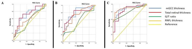
The study included 9 healthy individuals, 18 patients with ocular hypertension, 28 with preperimetric glaucoma, and 31 with early glaucoma. Total retinal thickness, mGCC thickness, RNFLT, and G/T ratio were highest in the healthy group and decreased progressively in patients with ocular hypertension, preperimetric glaucoma, and early glaucoma. All comparisons between the groups were significant for these parameters (p<0.001 for all). Average RNFLT, average GCC, and total retinal thickness showed consistently higher AUROC than G/T ratio in the differentiation between healthy individuals and patients with ocular hypertension, preperimetric glaucoma, and early glaucoma.
G/T ratio does not contribute to separation of ocular hypertension, preperimetric glaucoma, and early glaucoma patients from the healthy population. Compared to the other parameters investigated, G/T had lower diagnostic value.
评估在白种人群中,黄斑神经节细胞复合体-总视网膜厚度(G/T)比值的诊断准确性。
本横断面研究共纳入 86 例患者。患者分为 4 组:健康组;高眼压组;亚临床青光眼组;早期青光眼组。使用海德堡 HD 频域光学相干断层扫描仪(德国海德堡工程公司)测量每位患者随机一只眼的黄斑神经节细胞复合体(mGCC)厚度、总视网膜厚度和视网膜神经纤维层厚度(RNFLT)。G/T 比值(%)计算为(mGCC 厚度/总视网膜厚度)x100。通过受试者工作特征曲线(AUROC)下面积分析和固定特异性水平的敏感性评估,检查每个参数诊断青光眼的能力。使用独立样本 t 检验比较健康组与不同患者组之间的测量值。
本研究纳入了 9 名健康个体、18 名高眼压患者、28 名亚临床青光眼患者和 31 名早期青光眼患者。健康组的总视网膜厚度、mGCC 厚度、RNFLT 和 G/T 比值最高,随着高眼压、亚临床青光眼和早期青光眼患者病情进展而逐渐降低。各组间所有参数的比较均有统计学意义(所有 p<0.001)。在区分健康个体与高眼压、亚临床青光眼和早期青光眼患者时,平均 RNFLT、平均 GCC 和总视网膜厚度的 AUROC 始终高于 G/T 比值。
G/T 比值不能区分高眼压、亚临床青光眼和早期青光眼患者与健康人群。与其他研究参数相比,G/T 比值的诊断价值较低。