• 文献检索
  • 文档翻译
  • 深度研究
  • 学术资讯
  • Suppr Zotero 插件Zotero 插件
  • 邀请有礼
  • 套餐&价格
  • 历史记录
应用&插件
Suppr Zotero 插件Zotero 插件浏览器插件Mac 客户端Windows 客户端微信小程序
定价
高级版会员购买积分包购买API积分包
服务
文献检索文档翻译深度研究API 文档MCP 服务
关于我们
关于 Suppr公司介绍联系我们用户协议隐私条款
关注我们

Suppr 超能文献

核心技术专利:CN118964589B侵权必究
粤ICP备2023148730 号-1Suppr @ 2026

文献检索

告别复杂PubMed语法,用中文像聊天一样搜索,搜遍4000万医学文献。AI智能推荐,让科研检索更轻松。

立即免费搜索

文件翻译

保留排版,准确专业,支持PDF/Word/PPT等文件格式,支持 12+语言互译。

免费翻译文档

深度研究

AI帮你快速写综述,25分钟生成高质量综述,智能提取关键信息,辅助科研写作。

立即免费体验

高眼压症、视野检查前及早期青光眼的频域光学相干断层扫描参数评估

Evaluation of spectral domain optical coherence tomography parameters in ocular hypertension, preperimetric, and early glaucoma.

作者信息

Aydogan Tuğba, Akçay BetÜl İlkay Sezgin, Kardeş Esra, Ergin Ahmet

机构信息

Department of Glaucoma, Eye Clinic, Ümraniye Training and Research Hospital, Ümraniye, Istanbul 34766, Turke.

出版信息

Indian J Ophthalmol. 2017 Nov;65(11):1143-1150. doi: 10.4103/ijo.IJO_157_17.

DOI:10.4103/ijo.IJO_157_17
PMID:29133640
原文链接:https://pmc.ncbi.nlm.nih.gov/articles/PMC5700582/
Abstract

PURPOSE

The objective of this study is to evaluate the diagnostic ability of retinal nerve fiber layer (RNFL), macular, optic nerve head (ONH) parameters in healthy subjects, ocular hypertension (OHT), preperimetric glaucoma (PPG), and early glaucoma (EG) patients, to reveal factors affecting the diagnostic ability of spectral domain-optical coherence tomography (SD-OCT) parameters and risk factors for glaucoma.

METHODS

Three hundred and twenty-six eyes (89 healthy, 77 OHT, 94 PPG, and 66 EG eyes) were analyzed. RNFL, macular, and ONH parameters were measured with SD-OCT. The area under the receiver operating characteristic curve (AUC) and sensitivity at 95% specificity was calculated. Logistic regression analysis was used to determine the glaucoma risk factors. Receiver operating characteristic regression analysis was used to evaluate the influence of covariates on the diagnostic ability of parameters.

RESULTS

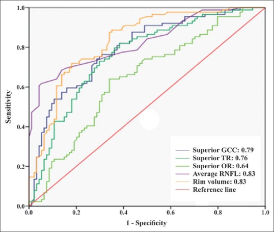
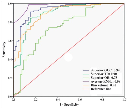
In PPG patients, parameters that had the largest AUC value were average RNFL thickness (0.83) and rim volume (0.83). In EG patients, parameter that had the largest AUC value was average RNFL thickness (0.98). The logistic regression analysis showed average RNFL thickness was a risk factor for both PPG and EG. Diagnostic ability of average RNFL and average ganglion cell complex thickness increased as disease severity increased. Signal strength index did not affect diagnostic abilities. Diagnostic ability of average RNFL and rim area increased as disc area increased.

CONCLUSION

When evaluating patients with glaucoma, patients at risk for glaucoma, and healthy controls RNFL parameters deserve more attention in clinical practice. Further studies are needed to fully understand the influence of covariates on the diagnostic ability of OCT parameters.

摘要

目的

本研究的目的是评估视网膜神经纤维层(RNFL)、黄斑、视神经乳头(ONH)参数在健康受试者、高眼压症(OHT)、视野检查前青光眼(PPG)和早期青光眼(EG)患者中的诊断能力,以揭示影响频域光学相干断层扫描(SD - OCT)参数诊断能力的因素及青光眼的危险因素。

方法

对326只眼(89只健康眼、77只高眼压症眼、94只视野检查前青光眼眼和66只早期青光眼眼)进行分析。使用SD - OCT测量RNFL、黄斑和ONH参数。计算受试者操作特征曲线(AUC)下面积及95%特异性时的敏感度。采用逻辑回归分析确定青光眼危险因素。采用受试者操作特征回归分析评估协变量对参数诊断能力的影响。

结果

在视野检查前青光眼患者中,AUC值最大的参数是平均RNFL厚度(0.83)和视盘边缘体积(0.83)。在早期青光眼患者中,AUC值最大的参数是平均RNFL厚度(0.98)。逻辑回归分析显示,平均RNFL厚度是视野检查前青光眼和早期青光眼的危险因素。随着疾病严重程度增加,平均RNFL和平均神经节细胞复合体厚度的诊断能力增强。信号强度指数不影响诊断能力。随着视盘面积增加,平均RNFL和视盘边缘面积的诊断能力增强。

结论

在评估青光眼患者、青光眼高危患者和健康对照时,临床实践中RNFL参数值得更多关注。需要进一步研究以充分了解协变量对OCT参数诊断能力的影响。

https://cdn.ncbi.nlm.nih.gov/pmc/blobs/9941/5700582/c837c301a245/IJO-65-1143-g006.jpg
https://cdn.ncbi.nlm.nih.gov/pmc/blobs/9941/5700582/7da0f2095797/IJO-65-1143-g004.jpg
https://cdn.ncbi.nlm.nih.gov/pmc/blobs/9941/5700582/c837c301a245/IJO-65-1143-g006.jpg
https://cdn.ncbi.nlm.nih.gov/pmc/blobs/9941/5700582/7da0f2095797/IJO-65-1143-g004.jpg
https://cdn.ncbi.nlm.nih.gov/pmc/blobs/9941/5700582/c837c301a245/IJO-65-1143-g006.jpg

相似文献

1
Evaluation of spectral domain optical coherence tomography parameters in ocular hypertension, preperimetric, and early glaucoma.高眼压症、视野检查前及早期青光眼的频域光学相干断层扫描参数评估
Indian J Ophthalmol. 2017 Nov;65(11):1143-1150. doi: 10.4103/ijo.IJO_157_17.
2
Diagnostic Ability of Wide-field Retinal Nerve Fiber Layer Maps Using Swept-Source Optical Coherence Tomography for Detection of Preperimetric and Early Perimetric Glaucoma.使用扫频光学相干断层扫描技术的广角视网膜神经纤维层图谱对视野缺损前和早期视野缺损性青光眼的诊断能力
J Glaucoma. 2017 Jun;26(6):577-585. doi: 10.1097/IJG.0000000000000662.
3
Ganglion cell-inner plexiform layer thickness of high definition optical coherence tomography in perimetric and preperimetric glaucoma.高分辨率光学相干断层扫描在视野缺损性青光眼和早期青光眼患者中的神经节细胞-内丛状层厚度研究
Invest Ophthalmol Vis Sci. 2014 Jul 11;55(8):4768-75. doi: 10.1167/iovs.14-14598.
4
Application of optical coherence tomography in glaucoma suspect eyes.光学相干断层扫描在青光眼可疑眼中的应用。
Clin Exp Optom. 2012 Jan;95(1):78-88. doi: 10.1111/j.1444-0938.2011.00654.x. Epub 2011 Oct 10.
5
Evaluation of macular ganglion cell analysis compared to retinal nerve fiber layer thickness for preperimetric glaucoma diagnosis.评价黄斑神经节细胞分析与视网膜神经纤维层厚度在青光眼前期诊断中的比较。
Indian J Ophthalmol. 2018 Apr;66(4):511-516. doi: 10.4103/ijo.IJO_1039_17.
6
Detection of macular ganglion cell loss in preperimetric glaucoma patients with localized retinal nerve fibre defects by spectral-domain optical coherence tomography.利用频域光学相干断层扫描检测局限性视网膜神经纤维层缺损的周边视野正常青光眼患者的黄斑神经节细胞丢失。
Clin Exp Ophthalmol. 2013 Dec;41(9):870-80. doi: 10.1111/ceo.12142. Epub 2013 Jul 5.
7
Comparison of glaucoma-diagnostic ability between wide-field swept-source OCT retinal nerve fiber layer maps and spectral-domain OCT.宽视野扫频源光学相干断层扫描视网膜神经纤维层图与谱域光学相干断层扫描诊断青光眼能力的比较。
Eye (Lond). 2018 Sep;32(9):1483-1492. doi: 10.1038/s41433-018-0104-5. Epub 2018 May 23.
8
Comparison of different spectral domain OCT scanning protocols for diagnosing preperimetric glaucoma.不同谱域 OCT 扫描方案诊断青光眼前期的比较。
Invest Ophthalmol Vis Sci. 2013 May 13;54(5):3417-25. doi: 10.1167/iovs.13-11676.
9
Structure-function relationship and diagnostic value of RNFL Area Index compared with circumpapillary RNFL thickness by spectral-domain OCT.频域光相干断层扫描的神经纤维层面积指数与环周神经纤维层厚度的结构-功能关系及其诊断价值。
J Glaucoma. 2013 Feb;22(2):88-97. doi: 10.1097/IJG.0b013e318231202f.
10
Glaucoma Detection Ability of Macular Ganglion Cell-Inner Plexiform Layer Thickness in Myopic Preperimetric Glaucoma.黄斑神经节细胞-内丛状层厚度在近视性视野缺损前期青光眼患者中的青光眼检测能力
Invest Ophthalmol Vis Sci. 2015 Dec;56(13):8306-13. doi: 10.1167/iovs.15-18141.

引用本文的文献

1
Comparative evaluation of OCT with OCTA changes at the optic disc and macula in glaucoma suspect and early glaucoma.青光眼可疑患者和早期青光眼患者视盘和黄斑区光学相干断层扫描(OCT)与光学相干断层扫描血管造影(OCTA)变化的比较评估
Indian J Ophthalmol. 2025 Mar 1;73(Suppl 2):S260-S266. doi: 10.4103/IJO.IJO_2575_23. Epub 2024 Oct 25.
2
Retinal functional and structural changes in patients with Parkinson's disease.帕金森病患者的视网膜功能和结构变化。
BMC Neurol. 2023 Sep 18;23(1):330. doi: 10.1186/s12883-023-03373-6.
3
The Comparison of Age-related Change in Retinal Nerve Fiber Layer and Ganglion Cell Complex Thicknesses between Glaucoma Suspects and Healthy Individuals.

本文引用的文献

1
Comparison of diagnostic capability of macular ganglion cell complex and retinal nerve fiber layer among primary open angle glaucoma, ocular hypertension, and normal population using Fourier-domain optical coherence tomography and determining their functional correlation in Indian population.使用傅里叶域光学相干断层扫描比较原发性开角型青光眼、高眼压症和正常人群中黄斑神经节细胞复合体和视网膜神经纤维层的诊断能力,并确定它们在印度人群中的功能相关性。
Indian J Ophthalmol. 2016 Apr;64(4):296-302. doi: 10.4103/0301-4738.182941.
2
Ganglion cell-inner plexiform layer thickness of high definition optical coherence tomography in perimetric and preperimetric glaucoma.高分辨率光学相干断层扫描在视野缺损性青光眼和早期青光眼患者中的神经节细胞-内丛状层厚度研究
Invest Ophthalmol Vis Sci. 2014 Jul 11;55(8):4768-75. doi: 10.1167/iovs.14-14598.
3
青光眼可疑患者与健康个体视网膜神经纤维层及神经节细胞复合体厚度的年龄相关变化比较
J Curr Glaucoma Pract. 2023 Jan-Mar;17(1):22-29. doi: 10.5005/jp-journals-10078-1392.
4
First Description of Inheritance of a Postzygotic Mosaic Variant.首例合子后镶嵌变体遗传的描述。
Genes (Basel). 2022 Mar 8;13(3):478. doi: 10.3390/genes13030478.
5
Measurements of OCT Angiography Complement OCT for Diagnosing Early Primary Open-Angle Glaucoma.OCT 血管造影测量对早期原发性开角型青光眼的诊断有 OCT 的补充作用。
Ophthalmol Glaucoma. 2022 May-Jun;5(3):262-274. doi: 10.1016/j.ogla.2021.09.012. Epub 2021 Oct 9.
6
Agreement between Compass Fundus Perimeter New Grid and 10-2 Testing Protocols for Detecting Central Visual Field Defects.新网格型 Compass 眼底周边视野计与 10-2 测试方案检测中心视野缺损的一致性。
Ophthalmol Glaucoma. 2022 Mar-Apr;5(2):179-187. doi: 10.1016/j.ogla.2021.07.006. Epub 2021 Jul 20.
7
Optical Coherence Tomography Evaluation of Peripapillary and Macular Structure Changes in Pre-perimetric Glaucoma, Early Perimetric Glaucoma, and Ocular Hypertension: A Systematic Review and Meta-Analysis.光学相干断层扫描评估视野缺损前青光眼、早期视野缺损青光眼和高眼压症患者视乳头周围及黄斑结构变化:一项系统评价和荟萃分析
Front Med (Lausanne). 2021 Jul 1;8:696004. doi: 10.3389/fmed.2021.696004. eCollection 2021.
8
Evaluation of spectral domain optical coherence tomography parameters in discriminating preperimetric glaucoma from high myopia.频域光学相干断层扫描参数在鉴别视野缺损前青光眼与高度近视中的评估
Int J Ophthalmol. 2019 Jan 18;12(1):58-65. doi: 10.18240/ijo.2019.01.09. eCollection 2019.
9
Macula Vessel Density and Thickness in Early Primary Open-Angle Glaucoma.早期原发性开角型青光眼的黄斑血管密度和厚度。
Am J Ophthalmol. 2019 Mar;199:120-132. doi: 10.1016/j.ajo.2018.11.012. Epub 2018 Nov 26.
10
The assessment of structural changes on optic nerve head and macula in primary open angle glaucoma and ocular hypertension.原发性开角型青光眼和高眼压症患者视神经乳头及黄斑区结构改变的评估
Int J Ophthalmol. 2018 Oct 18;11(10):1631-1637. doi: 10.18240/ijo.2018.10.09. eCollection 2018.
Comparative study of macular ganglion cell complex thickness measured by spectral-domain optical coherence tomography in healthy eyes, eyes with preperimetric glaucoma, and eyes with early glaucoma.利用光谱域光学相干断层扫描技术对健康眼、视野检查前青光眼患者眼及早期青光眼患者眼黄斑神经节细胞复合体厚度的比较研究。
Jpn J Ophthalmol. 2014 May;58(3):244-51. doi: 10.1007/s10384-014-0315-7. Epub 2014 Mar 11.
4
Effect of scan quality on diagnostic accuracy of spectral-domain optical coherence tomography in glaucoma.扫描质量对频域光学相干断层扫描诊断青光眼准确性的影响。
Am J Ophthalmol. 2014 Mar;157(3):719-27.e1. doi: 10.1016/j.ajo.2013.12.012. Epub 2013 Dec 15.
5
Evaluation of retinal nerve fiber layer thickness measurements for glaucoma detection: GDx ECC versus spectral-domain OCT.评价用于青光眼检测的视网膜神经纤维层厚度测量:GDx ECC 与谱域 OCT。
J Glaucoma. 2014 Apr-May;23(4):232-9. doi: 10.1097/IJG.0b013e3182741afc.
6
Comparison of different spectral domain OCT scanning protocols for diagnosing preperimetric glaucoma.不同谱域 OCT 扫描方案诊断青光眼前期的比较。
Invest Ophthalmol Vis Sci. 2013 May 13;54(5):3417-25. doi: 10.1167/iovs.13-11676.
7
Relationship between macular ganglion cell complex thickness and macular outer retinal thickness: a spectral-domain optical coherence tomography study.黄斑神经节细胞复合体厚度与黄斑外层视网膜厚度的关系:频域光学相干断层扫描研究。
Clin Exp Ophthalmol. 2013 Sep-Oct;41(7):674-82. doi: 10.1111/ceo.12089. Epub 2013 Apr 11.
8
Comparison of ganglion cell and retinal nerve fiber layer thickness in primary open-angle glaucoma and normal tension glaucoma with spectral-domain OCT.应用谱域光学相干断层扫描比较原发性开角型青光眼和正常眼压型青光眼的神经节细胞和视网膜神经纤维层厚度。
Graefes Arch Clin Exp Ophthalmol. 2013 Mar;251(3):831-8. doi: 10.1007/s00417-012-2114-5. Epub 2012 Aug 18.
9
The applicability of ganglion cell complex parameters determined from SD-OCT images to detect glaucomatous eyes.从 SD-OCT 图像中确定的神经节细胞复合体参数检测青光眼眼的适用性。
J Glaucoma. 2013 Dec;22(9):713-8. doi: 10.1097/IJG.0b013e318259b2e1.
10
Effect of spectrum bias on the diagnostic accuracy of spectral-domain optical coherence tomography in glaucoma.光谱偏倚对频域光学相干断层扫描诊断青光眼准确性的影响。
Invest Ophthalmol Vis Sci. 2012 Feb 29;53(2):1058-65. doi: 10.1167/iovs.11-8463.